

Steven Gabel, MD, FACS
 Elite Coalition Physician
-
Posts
328 -
Joined
-
Last visited
Content Type
Forums
Profiles
Store
Gallery
Articles
Blogs
Events
Downloads
Posts posted by Steven Gabel, MD, FACS
-
-
This 48-year-old patient of mine had surgery 2 years ago with about 3900 grafts to the frontal hairline and mid-scalp regions. He recently contacted us and sent in a photo of himself on vacation (only 1 after photo at this time). He is extremely excited about the transformation that took place with his hair and appearance, and stated that it really improved his overall confidence.
He will be coming in soon for an appointment. I will present the full range of before and after photographs from the office when he follows up, hopefully in the near future.


-
I appreciate the positive comments about the hair transplant; in terms of his hair style, he usually grows it longer, but since it will take time for the crown to catch up to the sides, he is trying out a new style. Usually those "wings" are in a ponytail and he was kind enough to let us take it down for the photographs and video. (The rubber band that holds it together is actually seen on my assistants finger during the video).
Out of respect for the patient, I would ask that we focus on the results of the transplant, and how happy he is with the growth. When he puts the hair in a ponytail, we are not able to see the results as clearly - that is why I asked him to take it down. Once everything is grown out, I'll take some photos of his final hair style.
Thanks.
-
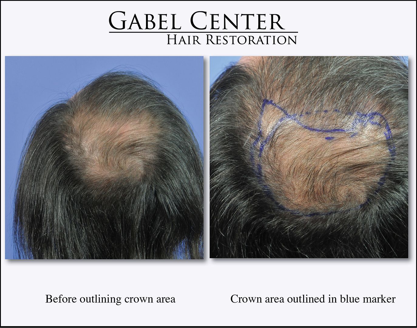
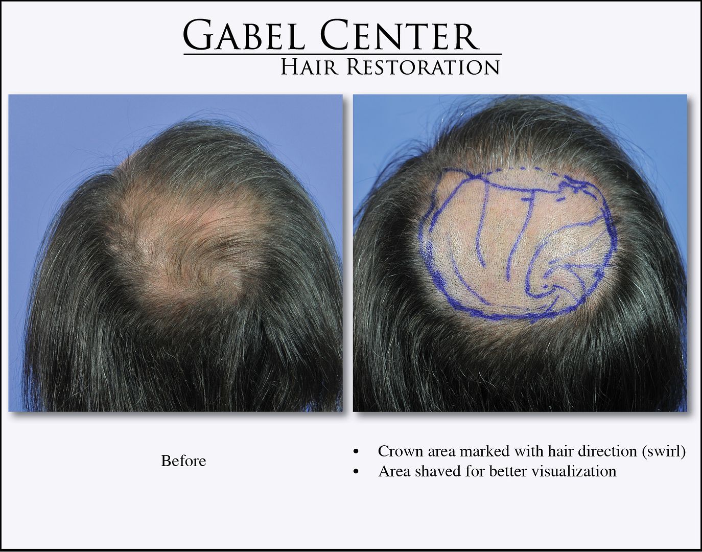
This 53-year-old gentleman started thinning years ago at the top and crown areas of his scalp. He had tried medications without any significant growth.
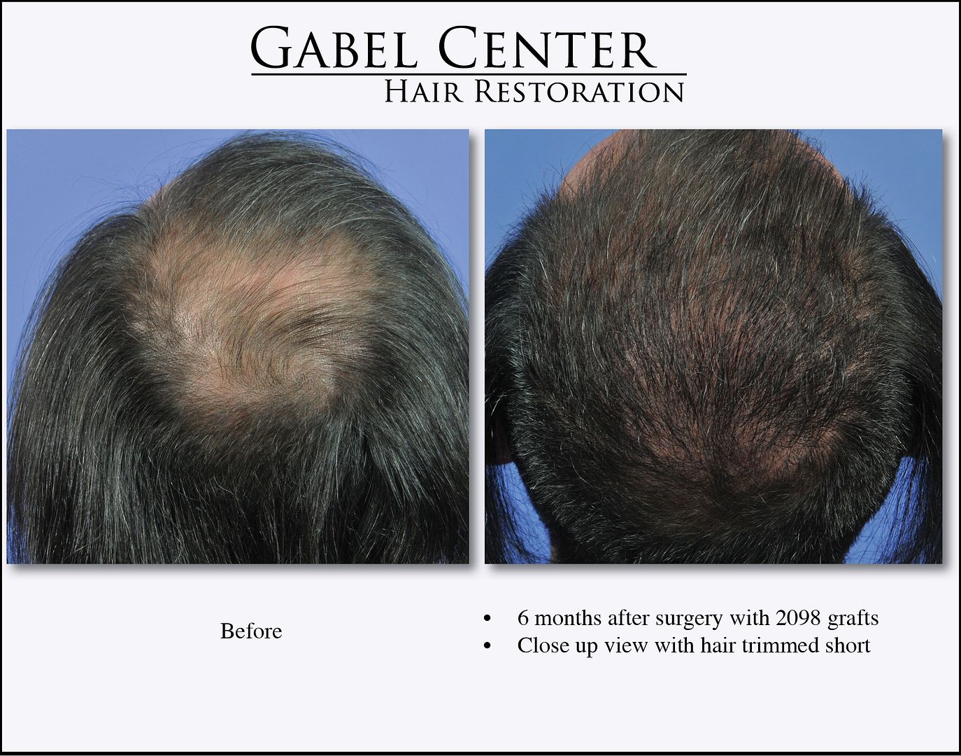
We transplanted 2098 grafts into the crown approximately 6 months ago. The series of photographs show the scalp as he presented before any surgery or haircuts, another photograph depicting the area of transplant showing his natural swirl pattern with blue markings, and the 6-month preliminary results.
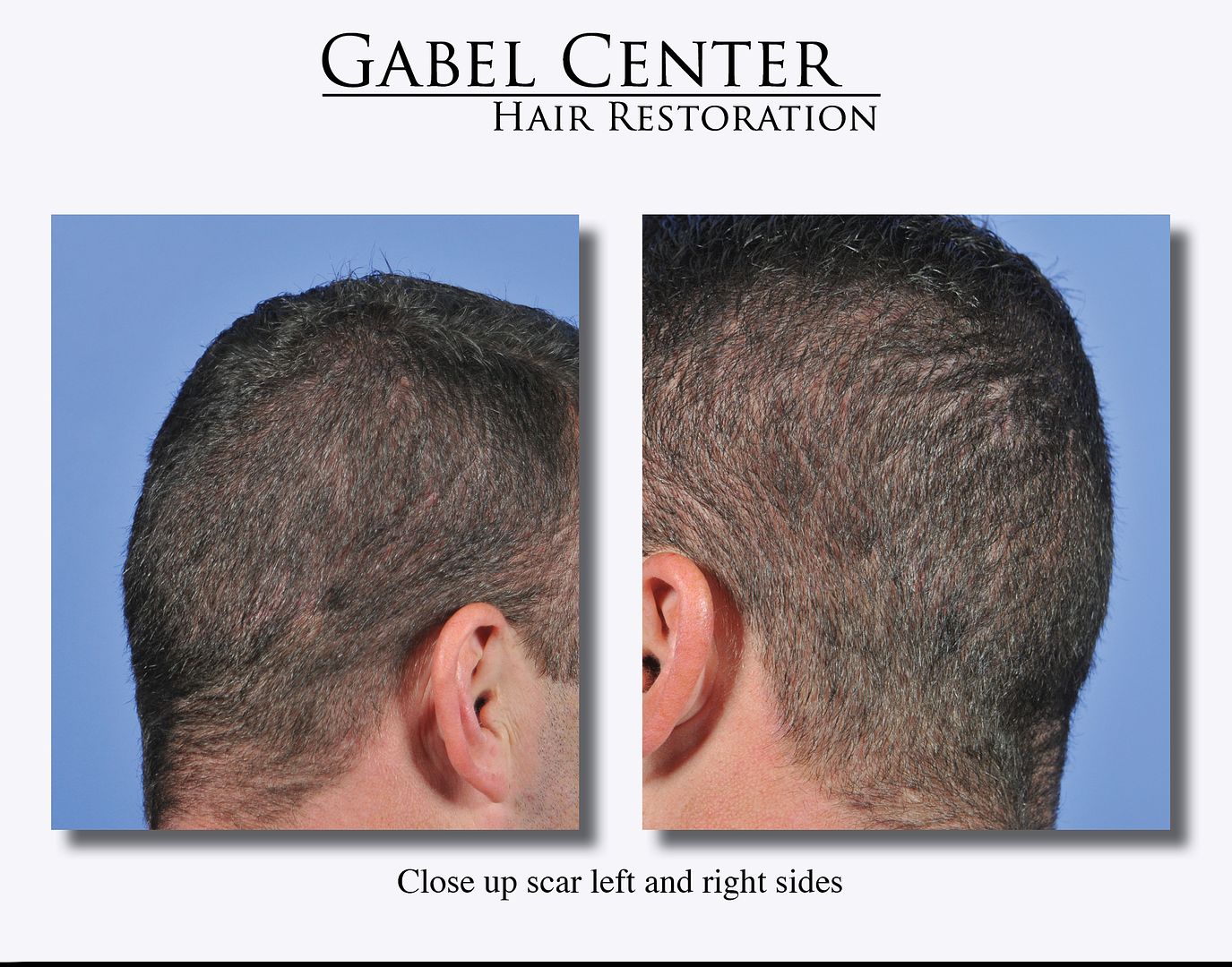
The swirl pattern was created by following some the “wispy” natural hairs that he had so I am able to make the slits in the natural direction and angle of his native hair. At 6 months, the transplanted follicular unit grafts are growing very well. He said that he did not shed that much after surgery and he has been already cutting his hair regularly. Also, the video shows the close up of the donor scar, which is thin and difficult to see even with his hair cut very short.
Overall the hair growth is excellent at this stage and we anticipate that the number of hairs will continue to increase and his hair will also thicken over time. He said he would come back in 3-month intervals to show his growth over time.
-
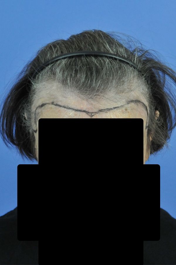
This patient is a 75-year-old female who had facial reconstructive surgery several years ago that left her with a high hairline and “too much forehead exposed.” To cover her forehead, 2279 grafts were placed to recreate her frontal hairline to lower it to a more natural position.
The photographs are 7 months after surgery. The hair is just starting to grow and already making a marked improvement. The frontal view is shown here; I will add several more photos in the near future. We look forward to posting her 1-year results when she returns.
-
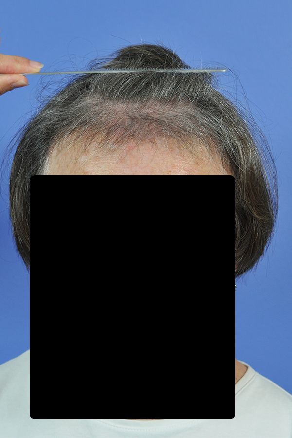
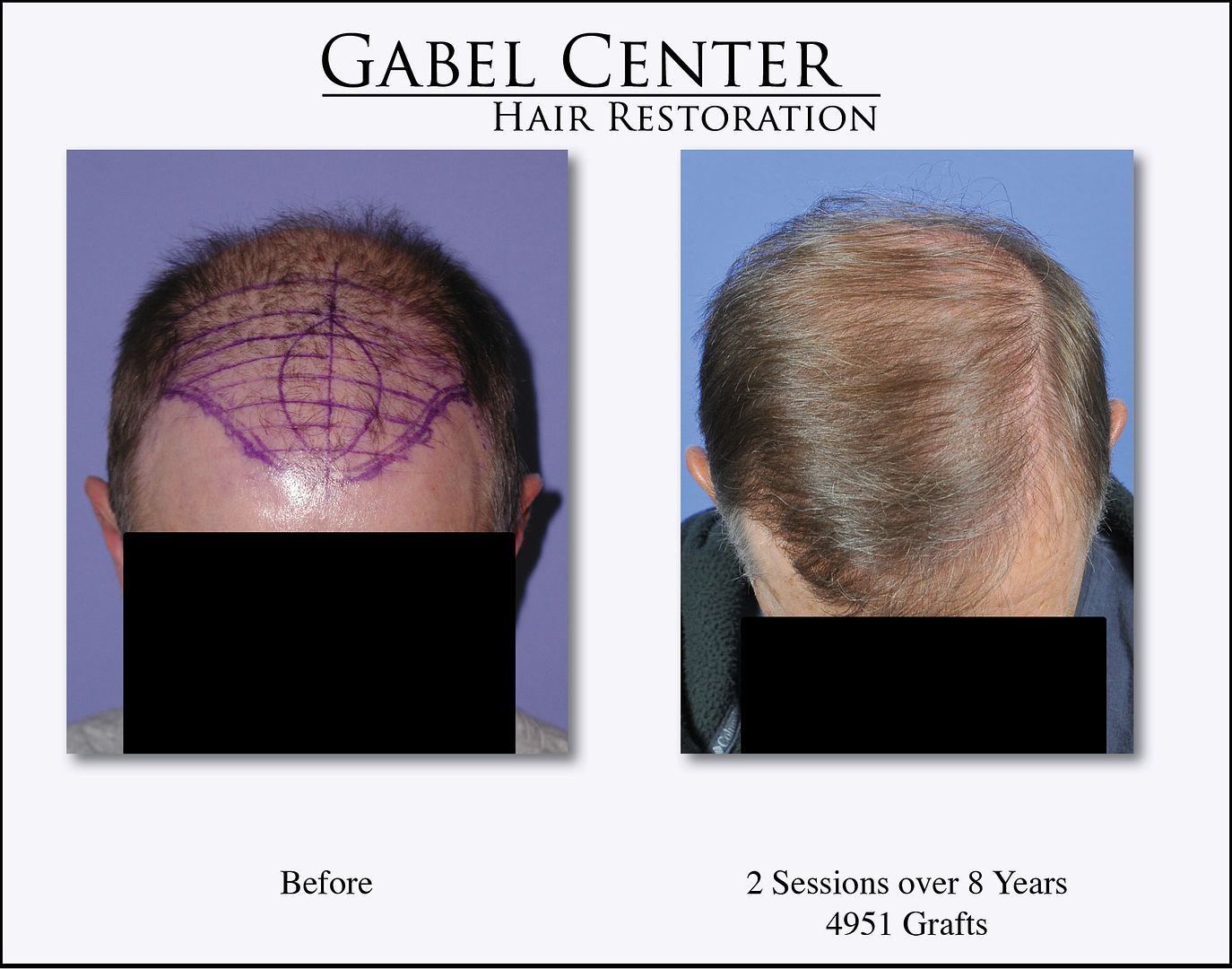
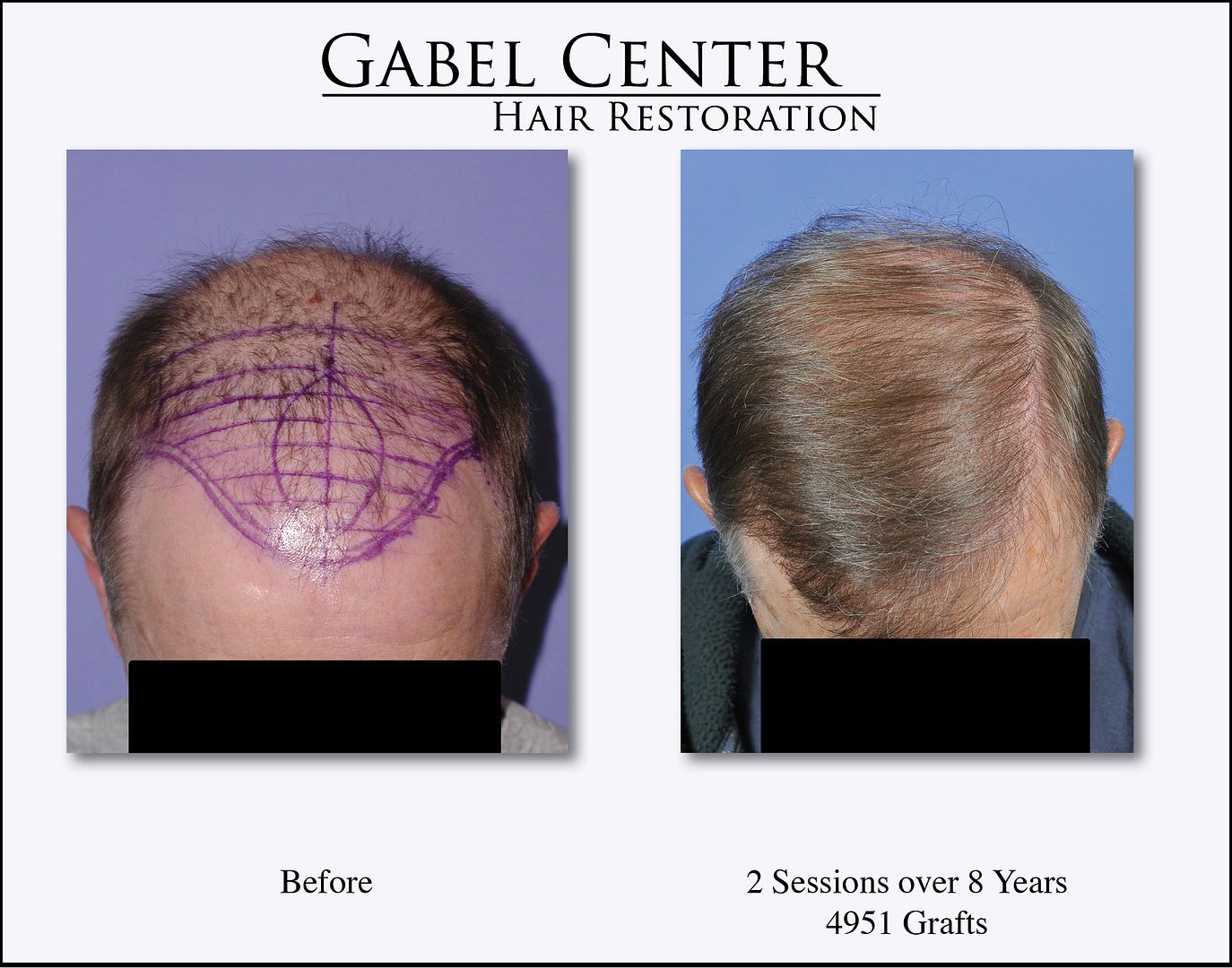
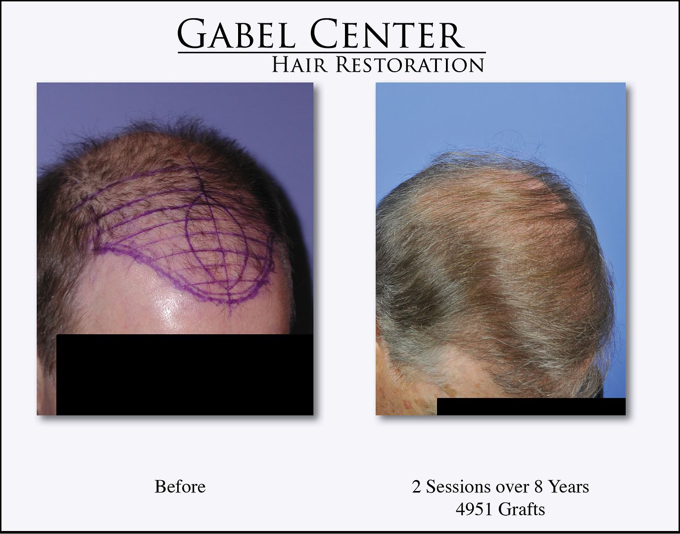
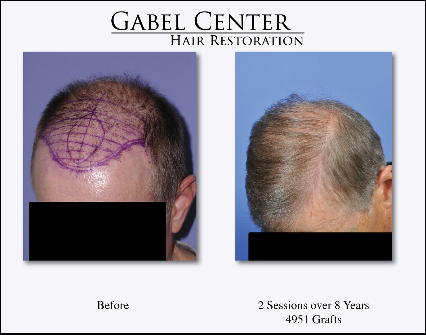
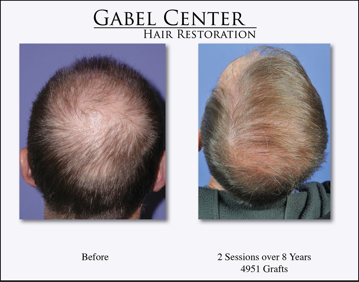
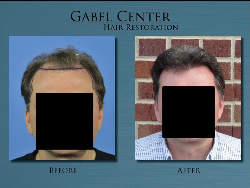
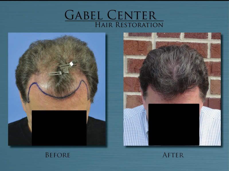
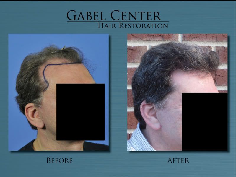
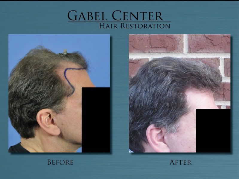
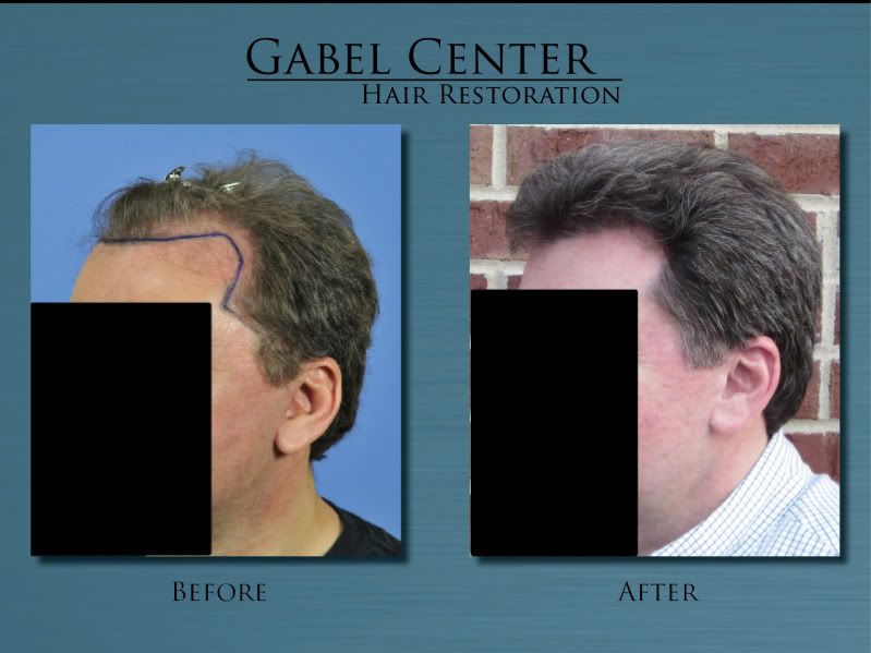
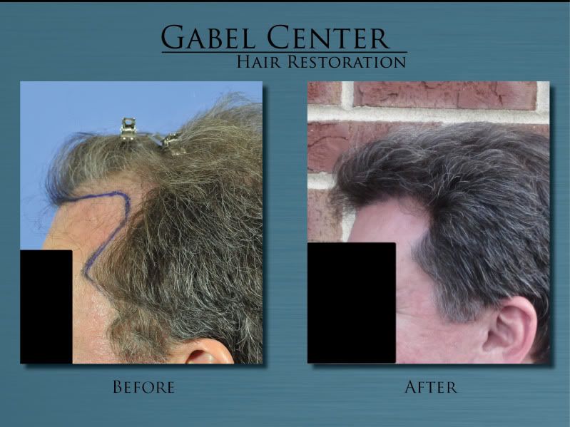
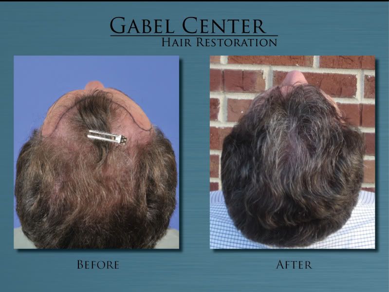
This patient is a 65-year-old gentleman who in 2005 had 2542 grafts placed into the frontal, mid-scalp/anterior crown region. Between 2005 and 2012, he had some more thinning of the native hairs and we added another 2409 grafts to the frontal and mid-scalp region, but this transplant, we added a good number of grafts into the crown. He is now 1 year out from his second transplant and is extremely happy with the result.
In 2005, I did not take the extensive preoperative and postoperative photographs that I now take so there are only a few direct comparisons for photographs. The gray/blond hair mixed with a few black hairs gives him a very natural appearance for his age and overall his goals were accomplished with the type of hair he has.
-
Othersyde
Here is the video of your first postoperative day. There are some nice close up shots of the hairline which I think you'll enjoy. Looking forward to watching how everything progresses.
-
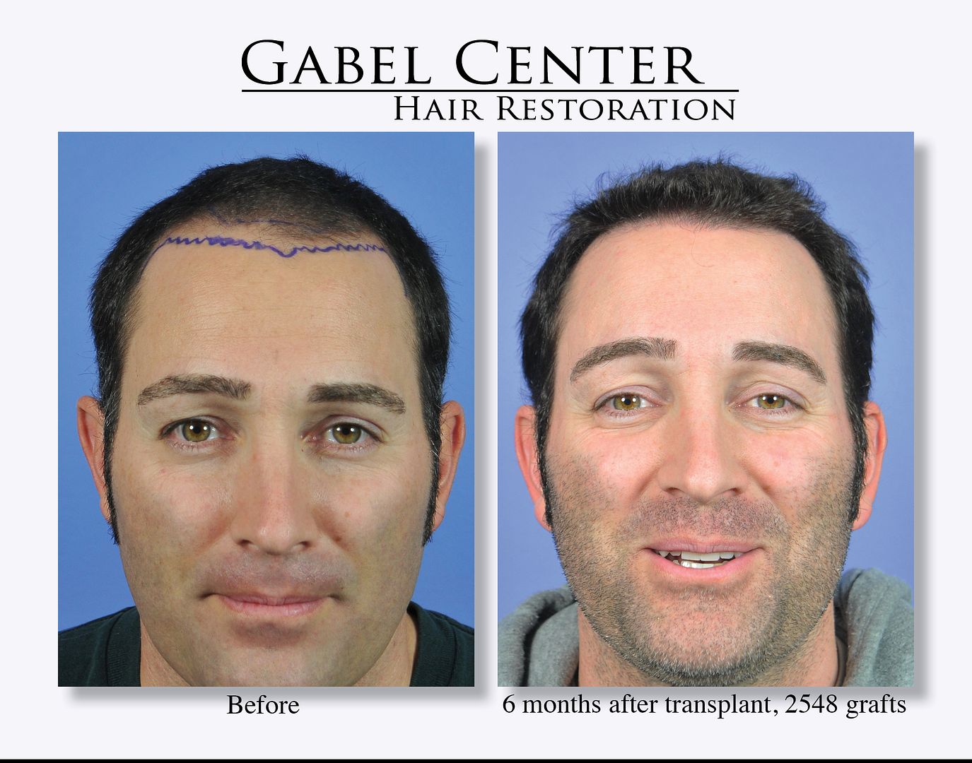
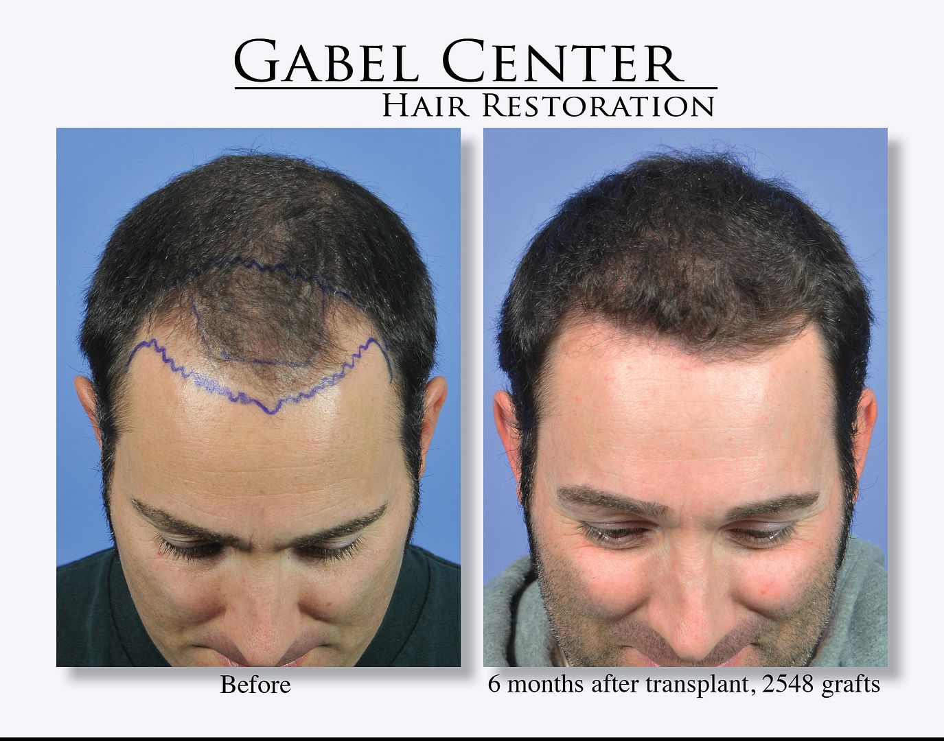
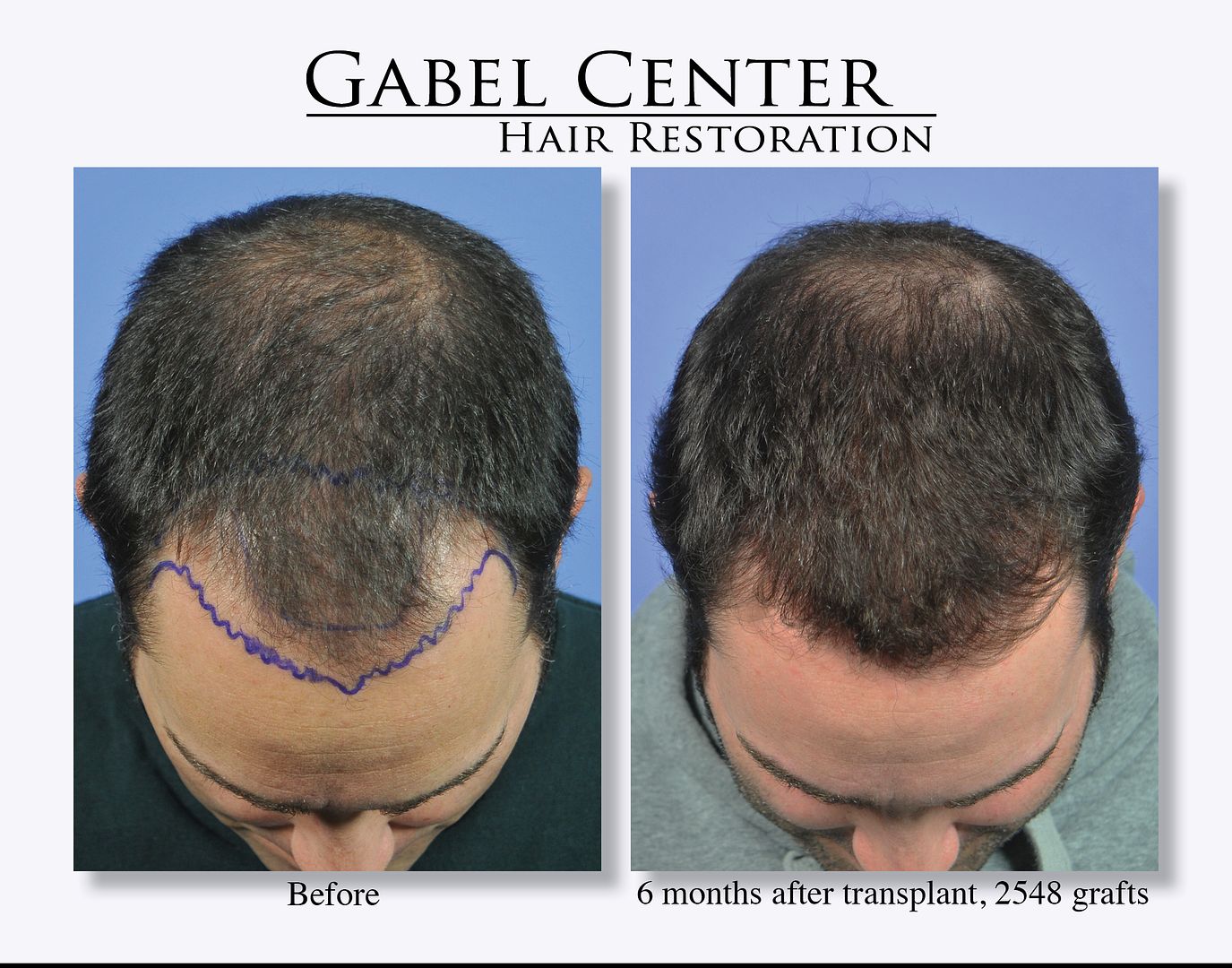
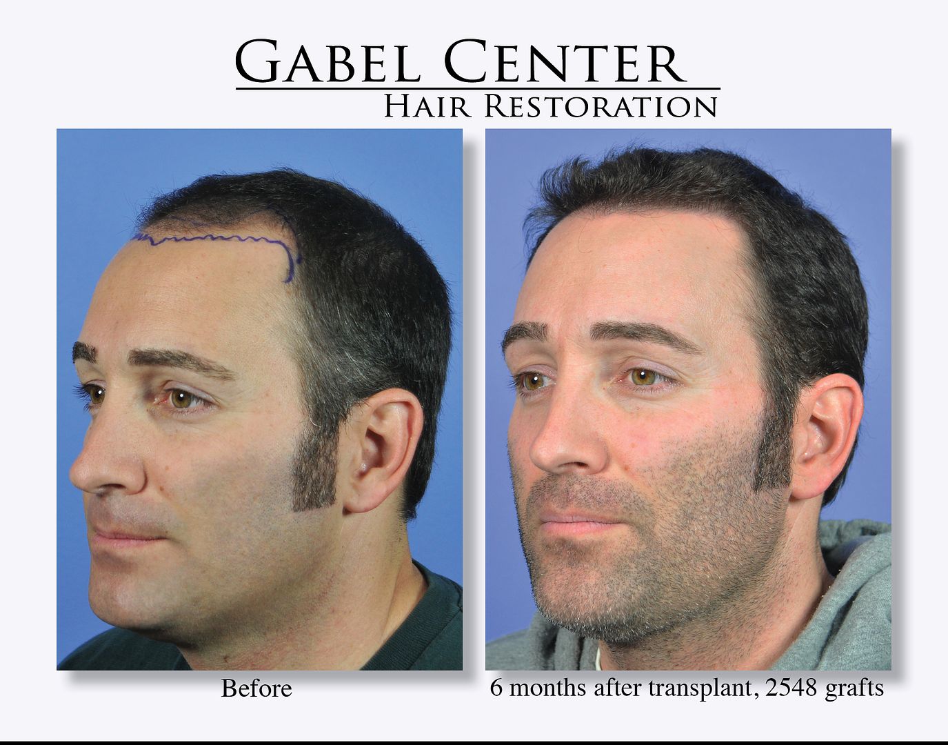
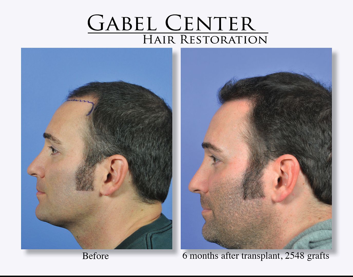
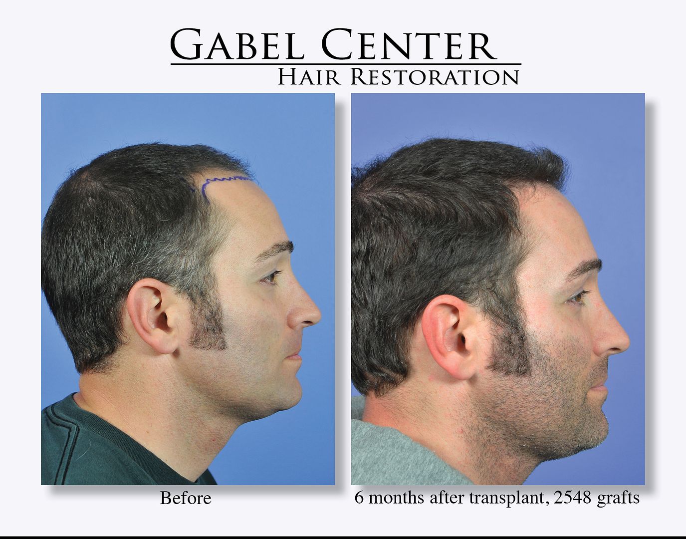
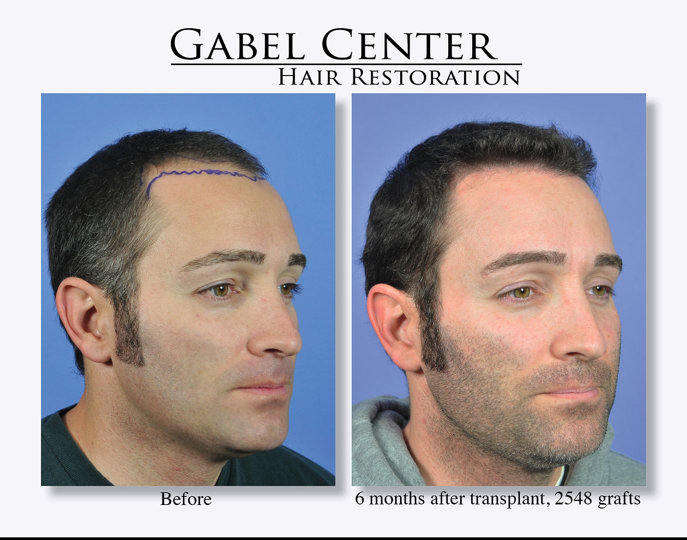
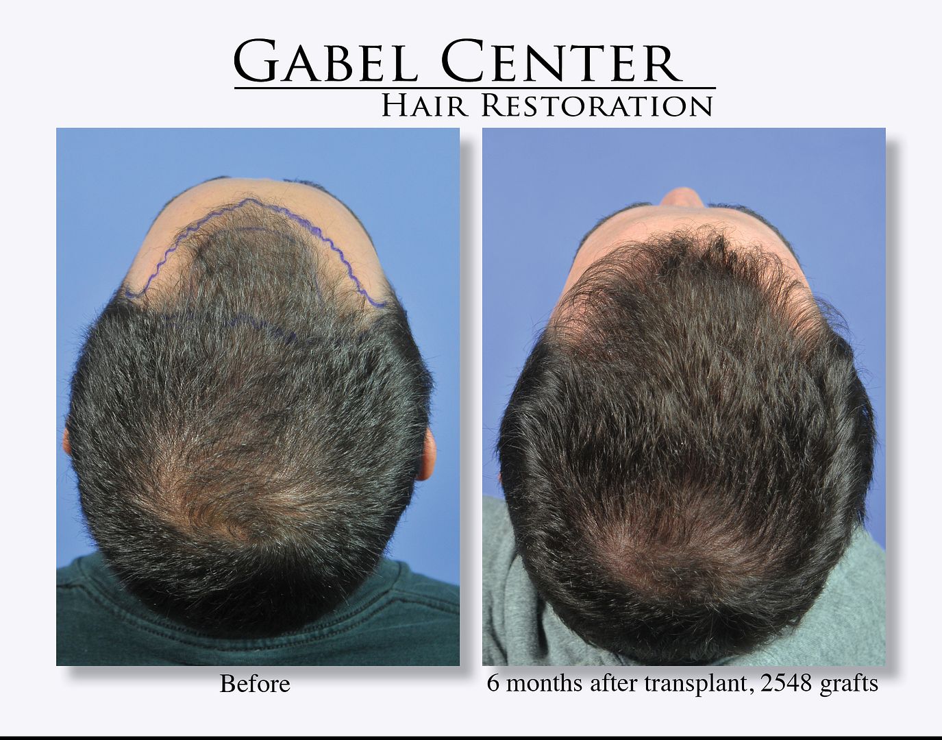
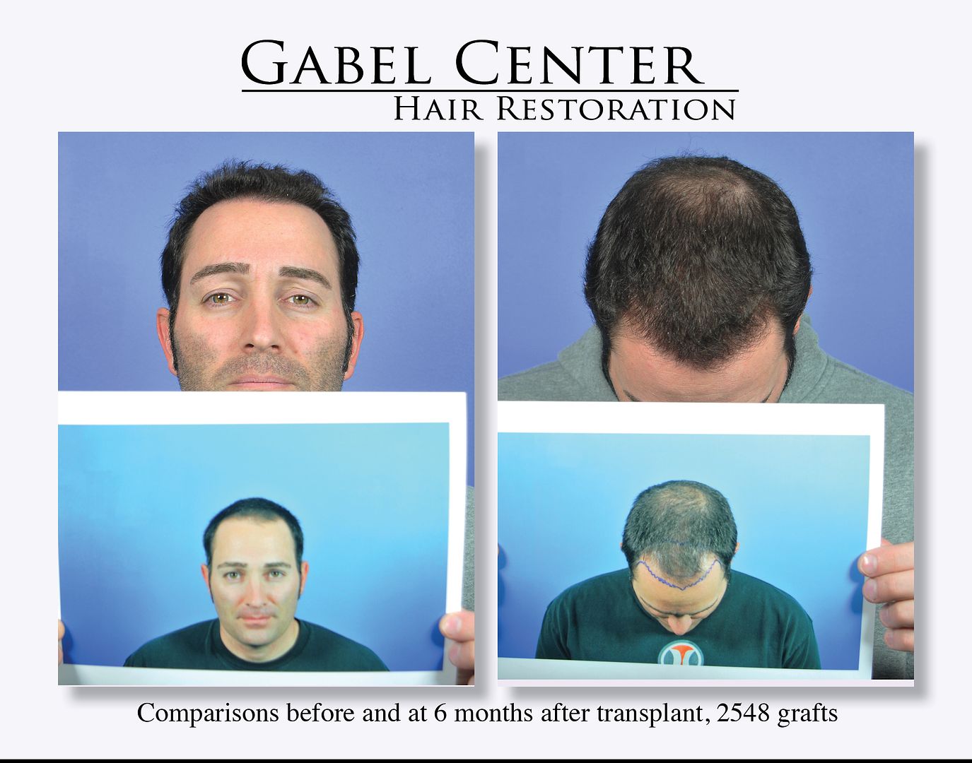
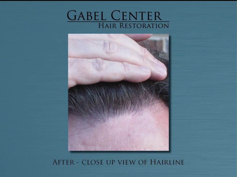
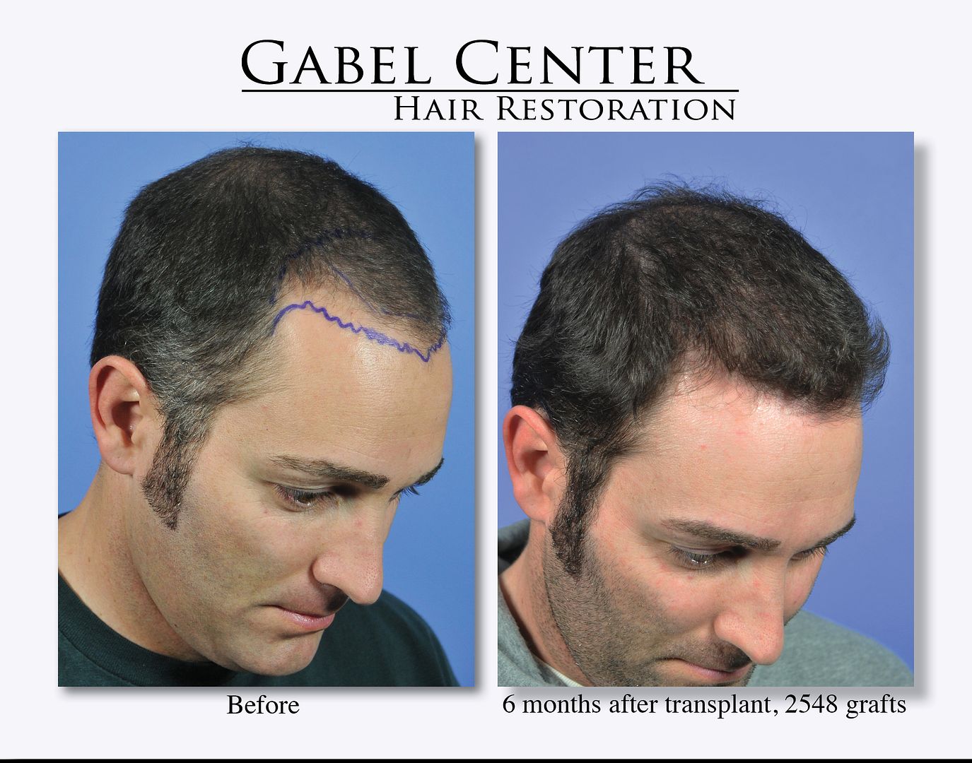
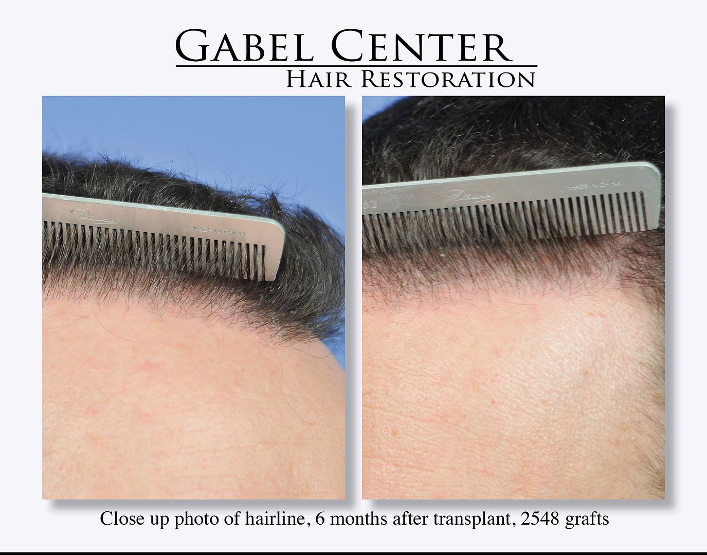
This patient is a 34-year-old gentleman who desired to thicken the frontal hairline and scalp. He was concerned also about the crown, but we elected to treat this area with medications at this point because of his age and see how well he responds.
He had 2548 grafts placed in the frontal hairline and frontal scalp 6 months ago. He stated that his hair started growing about 2 months ago and seen continued improvement each month. Since he is only 6 months out, his hair will continue to grow and thicken over the next 18 months.
-
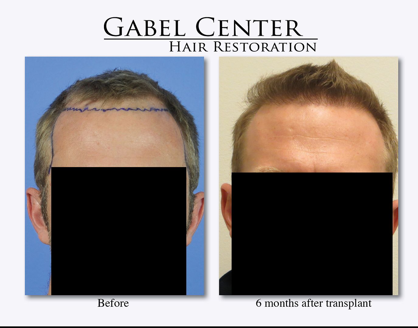
This patient had 2752 grafts placed into the frontal hairline and scalp. He is approximately 6 months out from his surgery and is starting to grow in. The grafts were placed in a coronal orientation and were dense packed. The photos were taken in our Seattle office so the background/camera are not the same as in our Oregon office. We will continue to update the photos as his hair grows in and thickens up.
-
I appreciate everyone’s patience while waiting for my response. My primary responsibility during the week is to the patients that I am operating on that day and I also wanted to make sure that I had spoken to this patient and obtained all the facts and his permission to openly discuss the case, which he has granted. I also asked him to send me additional photos, which I received yesterday.
First of all, as I said, I have been in touch with the patient and have had excellent phone conversations with him. Unfortunately, he lives in California while I am in Oregon so I am not able to evaluate him in person, but that will change this summer when I may be in his area; if that is not possible, we have made plans to see each other when I travel to San Francisco for the ISHRS meeting this fall. Either way, he was able to send some additional photos, which he took himself, which will be reviewed.
To get to the heart of the matter, the real question is: was this a failed hair transplant. To answer this question, we need to look objectively at the entire situation, not just a moment in time. The goals and realistic results must be fully discussed between the treating physician and the patient and it is the responsibility of the physician to convey what is realistically possible with both medical and surgical therapy. If the goals of the patient are not in line with what I feel is possible, then I will not treat that individual – that is a set up for failure.
I was contacted by this patient who desired more hair on his scalp. We had multiple phone consultations and a video consultation. At the time, he was able to tell me that his hair was thin, and he was not on any medications. We certainly discussed the pros/cons of medical therapy and that I am a big proponent of their use. In terms of the hair transplant, I discussed the fact that he is a NW Class VI with a large area to transplant. I stated multiple times that it would require more then 1 procedure to obtain a nice result. But, I also explained, and documented, that with a large surface area to transplant with thin hair, that the final results would not be dense, and it would have a thin appearance, and our goal would be to make it as natural as possible. He also inquired about the crown, which I recommended he wait until the frontal portion of the scalp was satisfactory.
When I was able to see the patient in person, indeed, he had thin hair, and a large area to transplant. Examination of the scalp revealed a higher percentage of smaller single and double follicular units as well. The proposed area to transplant was in excess of 150 square centimeters. See Figure 1. The goal of the first transplant was to place as many grafts as possible, safely, to cover the proposed area. During this consultation, which I have documented in 3 locations, I fully discussed the facts: 1. He has a large area to cover and the density on a first pass will be low. 2. His hair caliber is thin. 3. The follicular units bundles are smaller. 4. He will need several procedures to obtain the final result. All of these characteristics, I discussed in detail, will result in a thin appearance. And as I have documented in my consultation note, and I quote: “Discussed in detail expected results with fine hair.” Additionally, on my consent form, these are also spelled out in detail.
In terms of the transplant itself, it was uneventful. The graft breakdown was as follows:
Total: 3706 grafts
1’s: 1145 (31%) – Almost twice the normal value
2’s: 2127 (57%)
3’s: 423 (11%)
4’s: 11 (<1%)
88% 1 and 2 hair follicular units is a very high % of smaller haired grafts, which in turn, will yield a thinner result. In terms of the slit density, with such a large area, I was only able to obtain around 20 - 25 follicular units per sq. cm. That is less then 20% natural density. See figure 2 and figure 3 from this patient. Of note, when I perform a transplant going toward the crown, I will fade single hair follicular units into the crown region so it has a more natural appearance until the patient can return for a second procedure. I do not like the “wall” appearance that can result if I only put larger follicular units abutting the crown region if the crown is not transplanted. I do this because, even through we are staging the procedure, one never knows exactly when they will return for a second procedure and I want them to have a natural appearance between procedures. In his case, since we had so many smaller follicular units, it was not a problem to produce this.
Lastly, objectively looking at the 1 year result photos which he originally presented, they did not adequately show whether the grafts that I placed had grown or not. He said his hair was wet and he had product in it. And, frankly, I agree – they are not the best representation of a hair transplant. Therefore, I asked him to send me additional photos, after he washed his hair and removed any product, which he has presented. Again, I wish that I was able to take these with my professional camera and a macro lens for the best before and after comparison, but this is what I have to work with. If you look at Figure 4 (the photos sent to me by the patient – which he presented in an earlier thread), it appears to me that the transplanted grafts are growing. If you compare Figure 2 (the outline of the slits) and Figure 4 (the growing hairs), the area that was transplanted is growing well. So to answer question #2: did the actual hair transplant itself fail? The answer to that question is No. The hairs that I transplanted are growing, and according to the patient who I have spoken to on several occasions, he also feels that the transplanted area is growing.
To summarize: this patient has several characteristics, which ultimately gives a thinner appearance – low caliber hair thickness, a significant number of single/double follicular unit bundles (88%) and a large surface area to transplant, which ultimately gives a low density result. As discussed originally with this patient, he will need additional transplants to thicken up the frontal region to provide him with a higher density to satisfy his goals. In my opinion, this is not a failed transplant, but a work in progress. As his treating physician, I certainly empathize with his situation and will do whatever I can to continue to move forward to accomplish his goals.


-
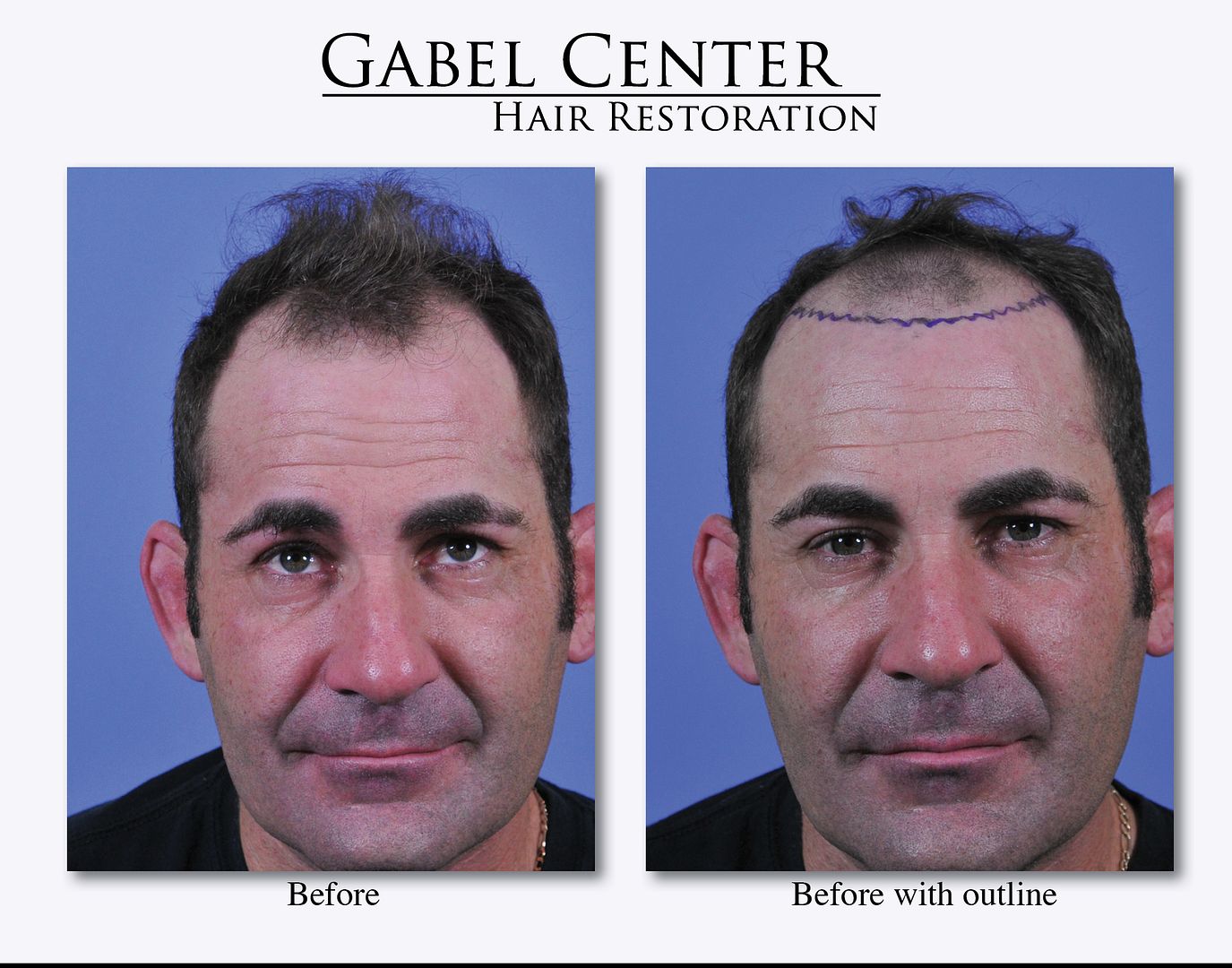
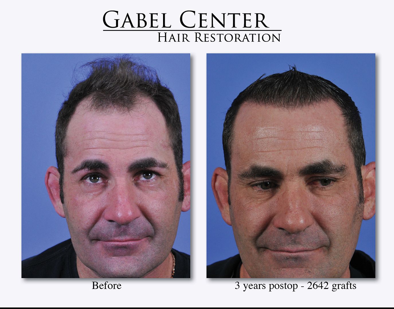
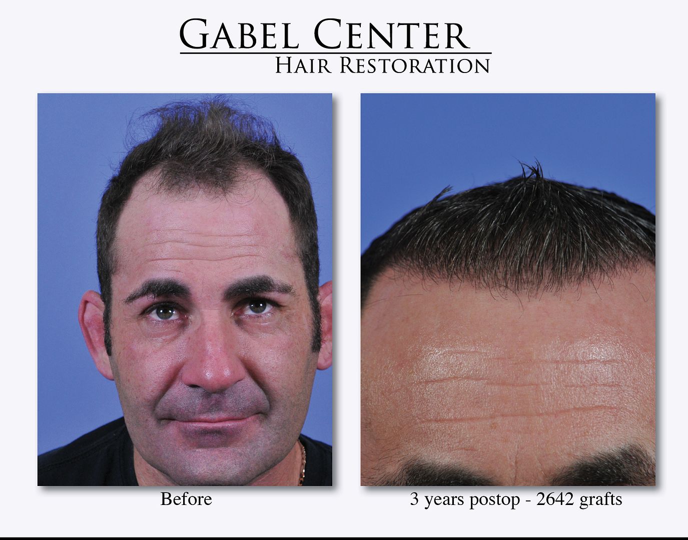
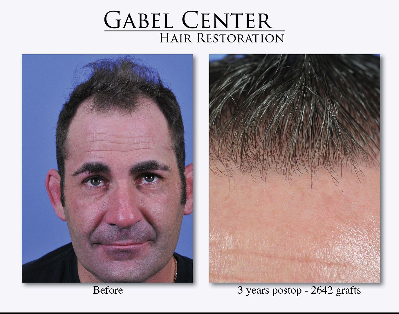
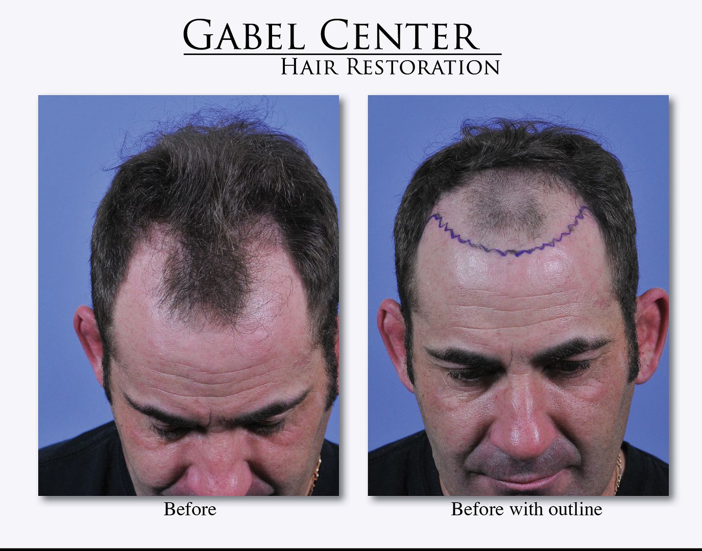
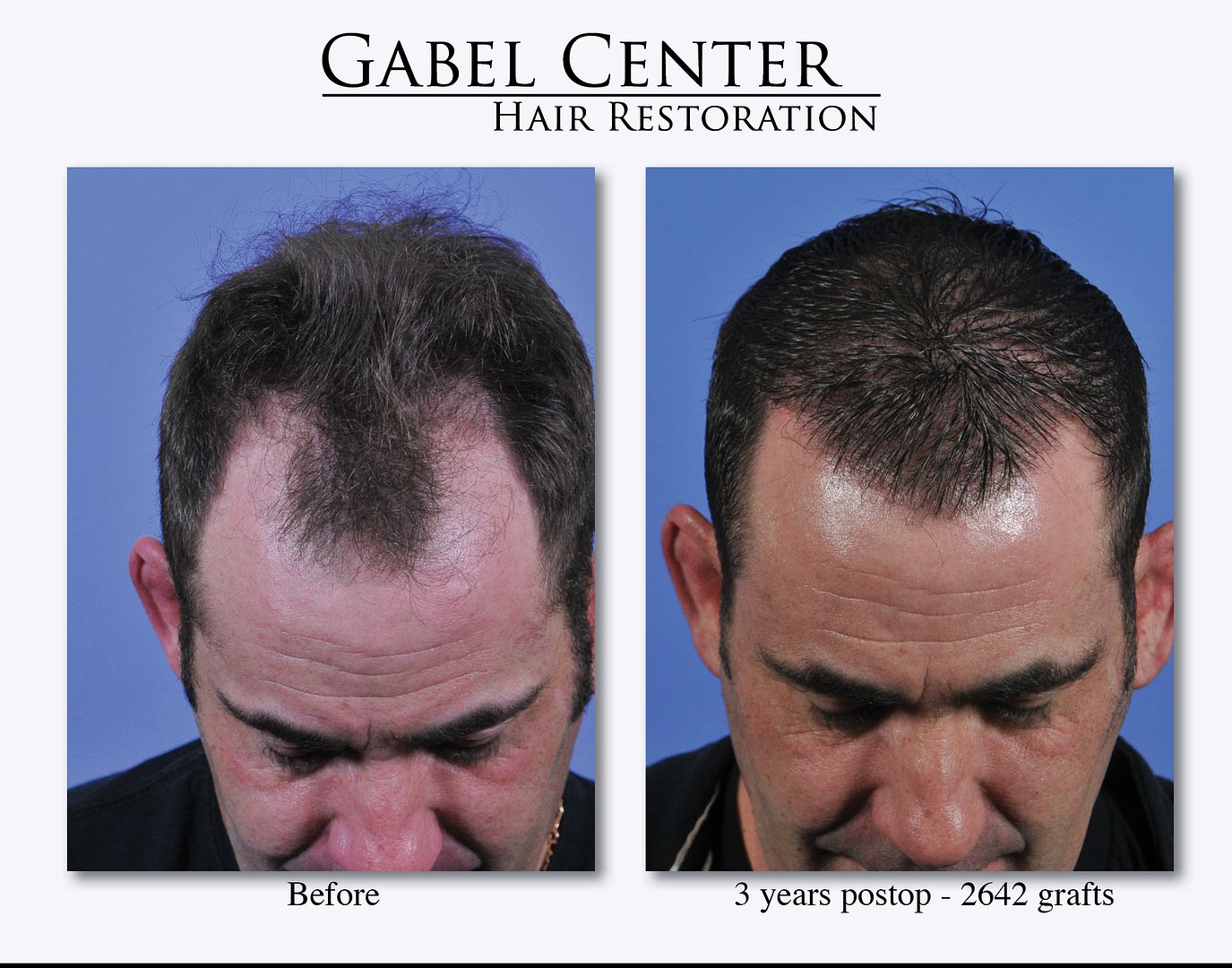
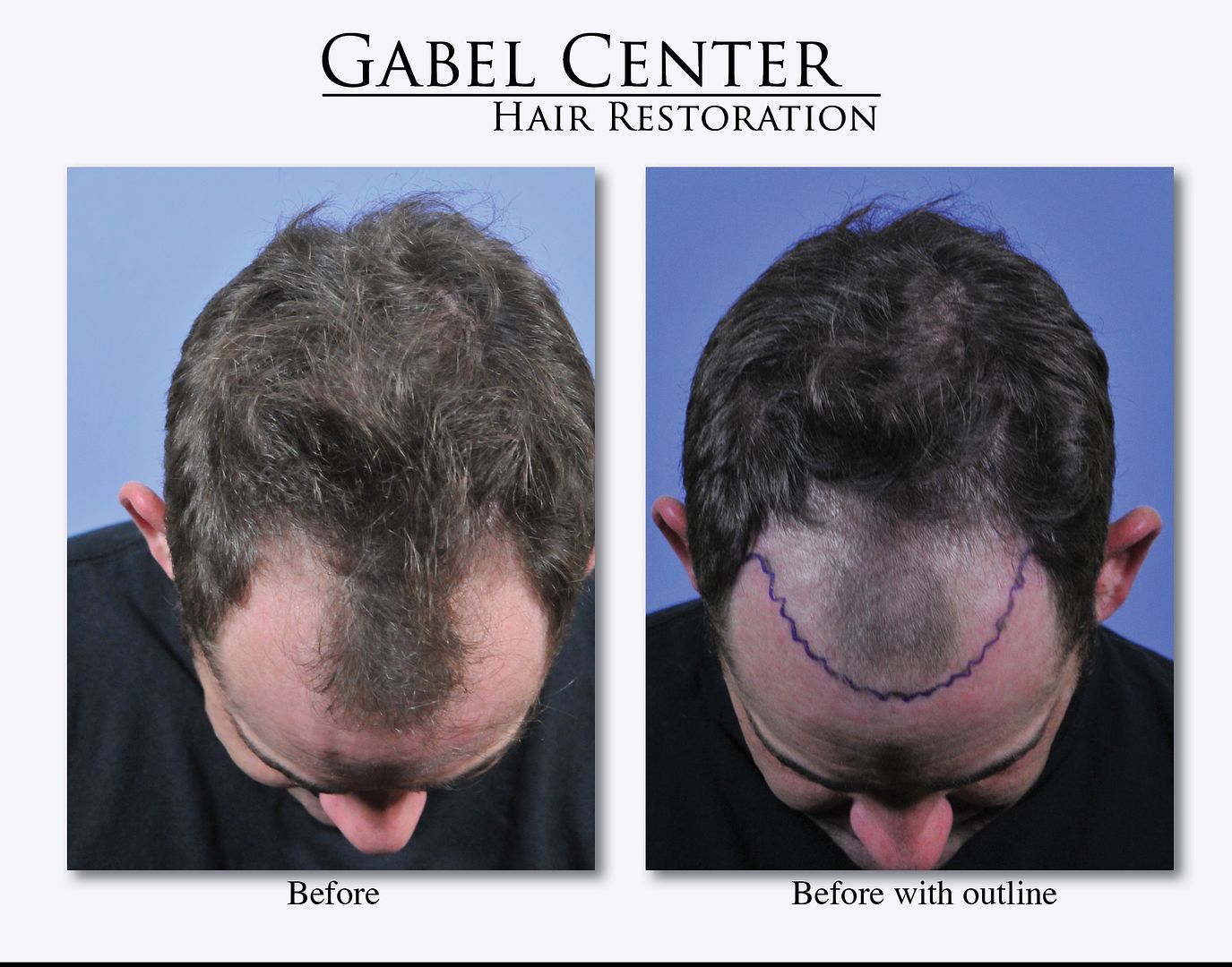
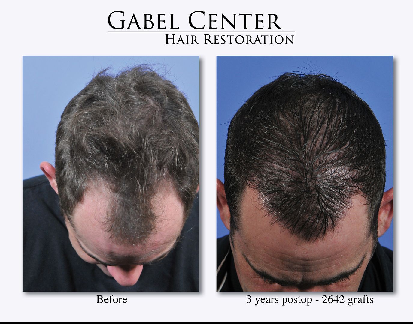
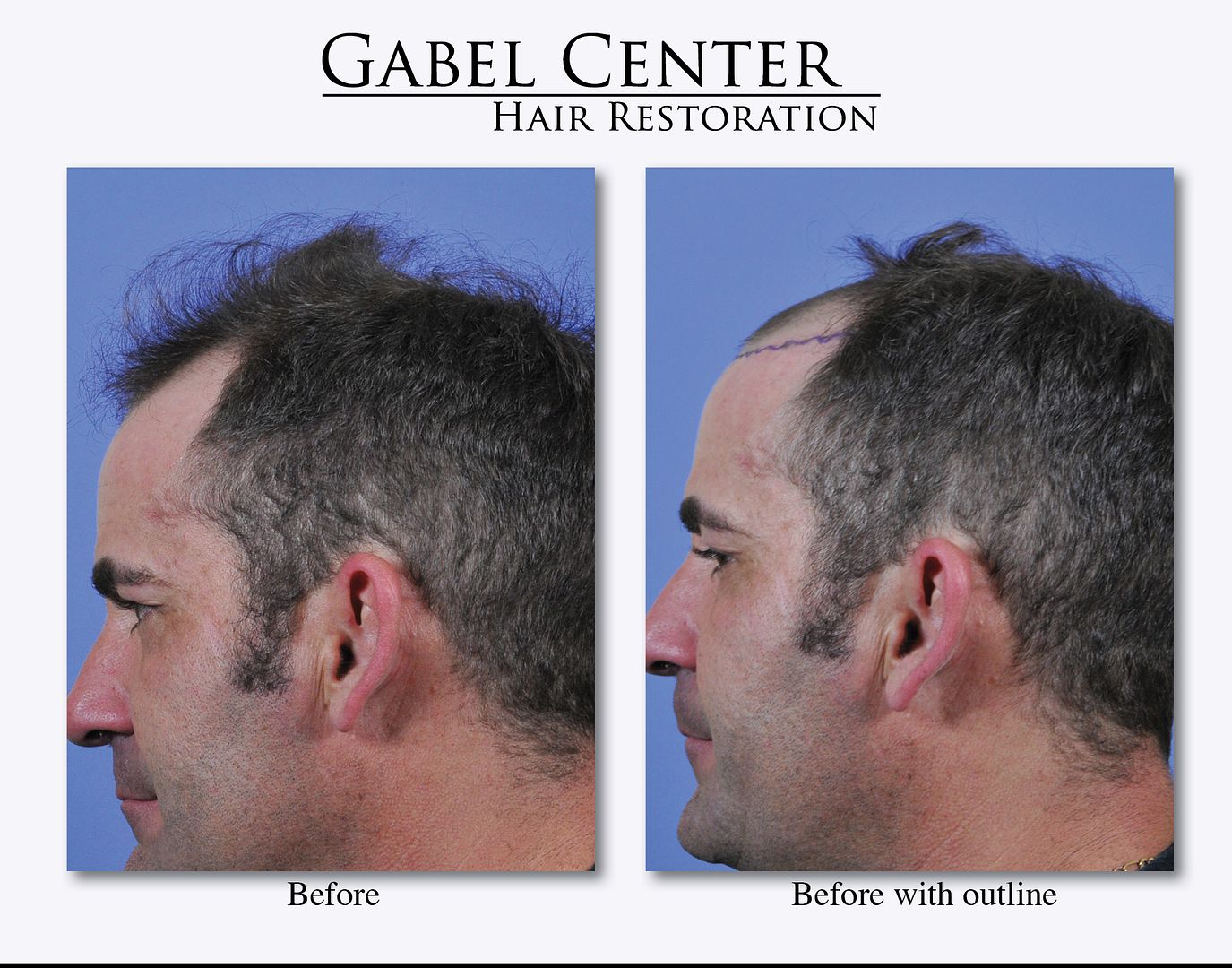
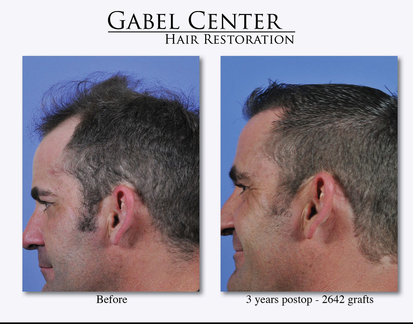
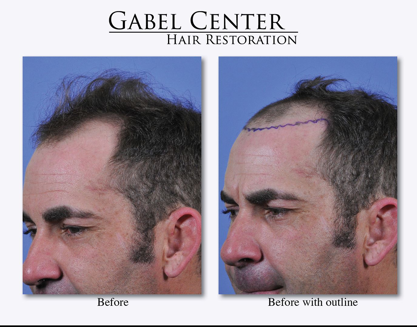
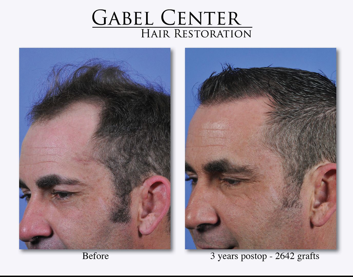
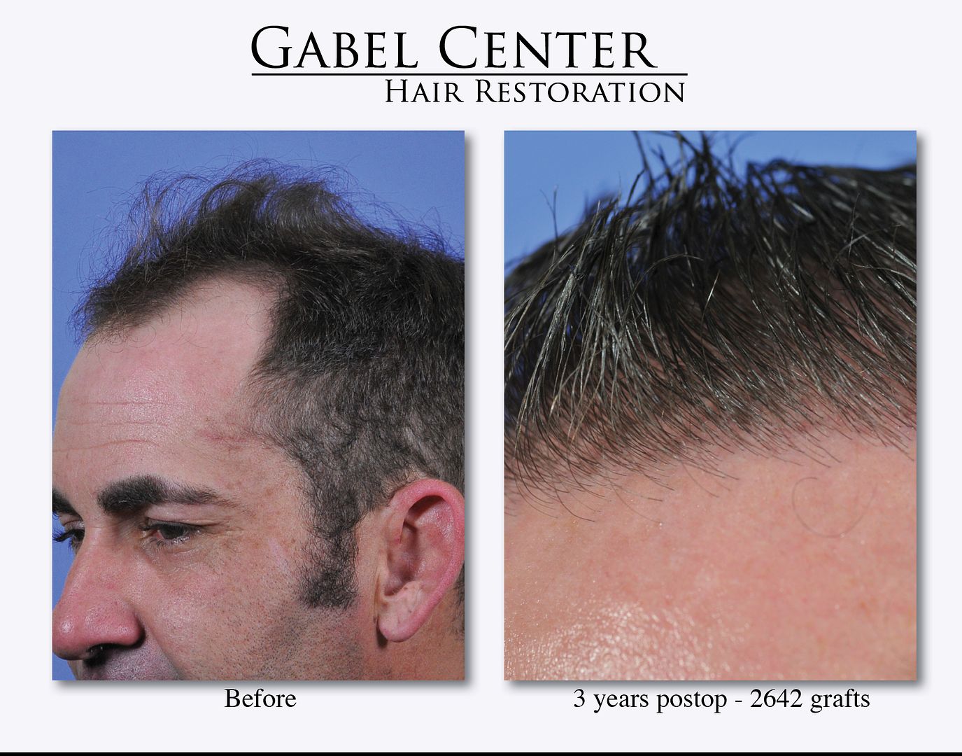
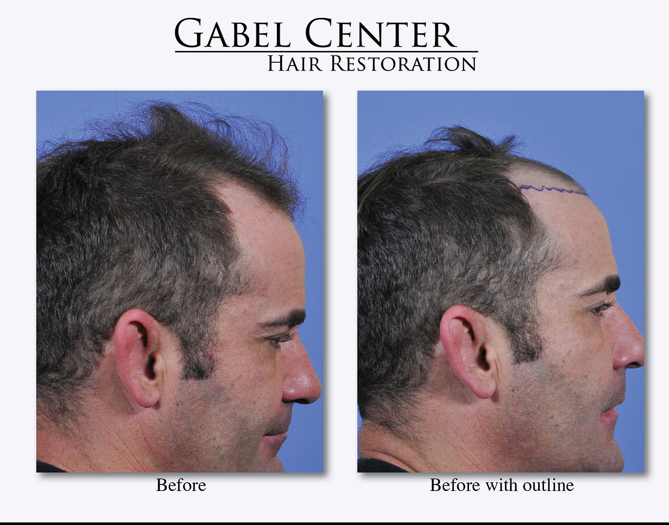
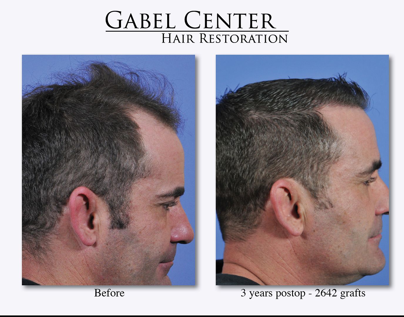
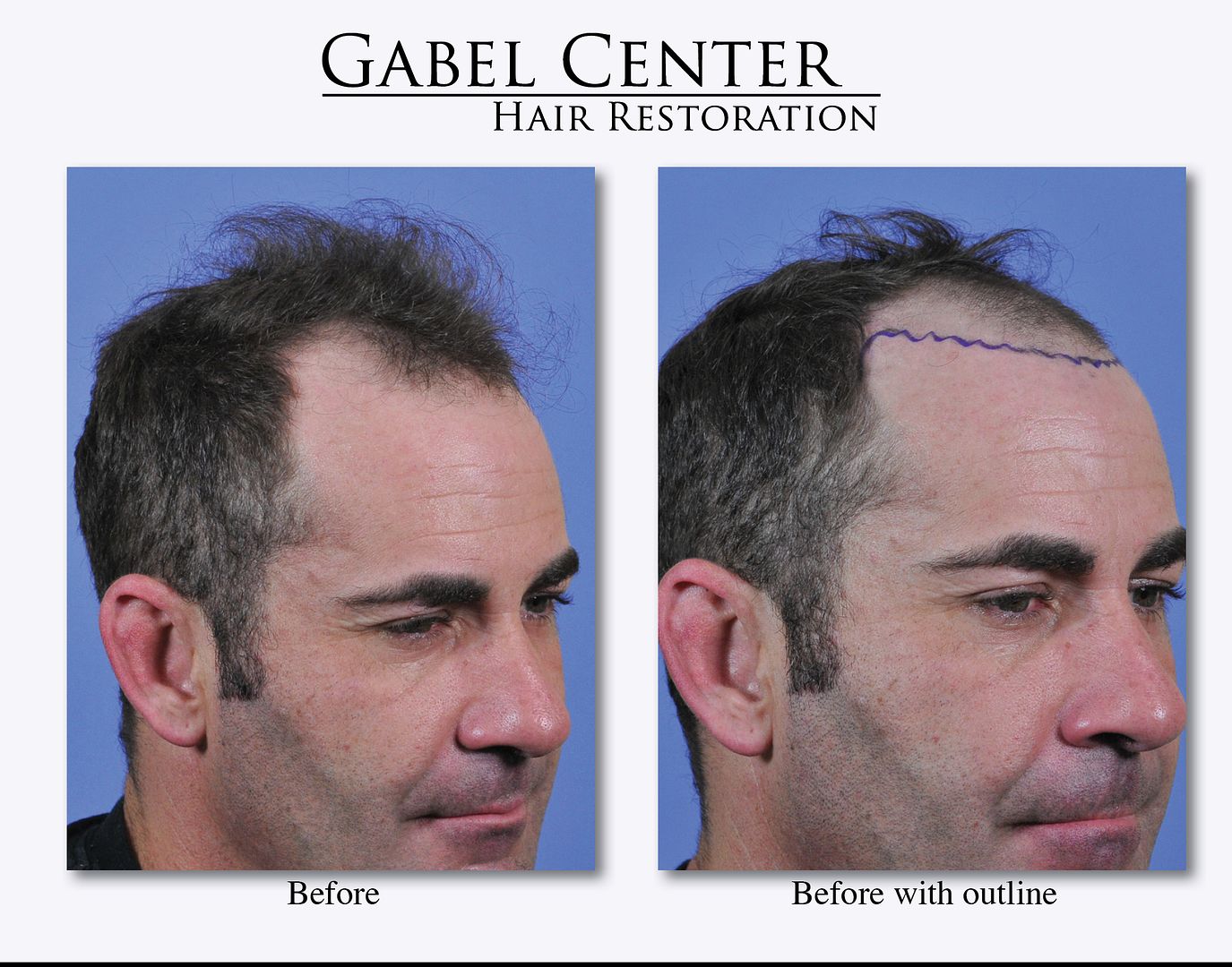
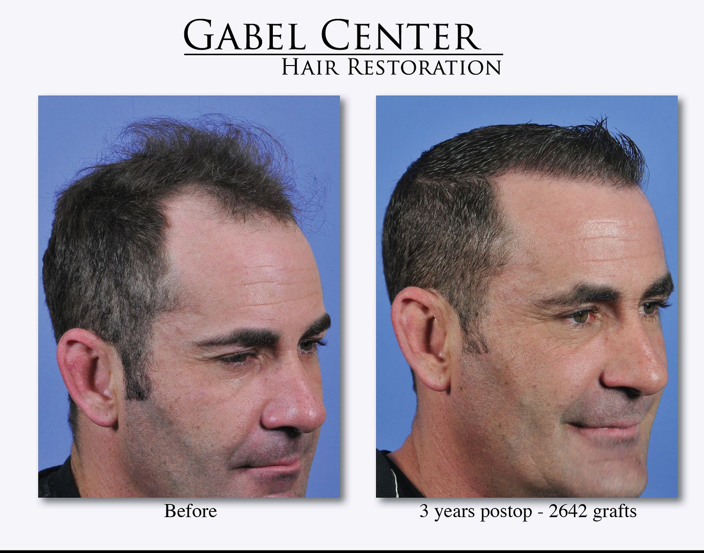
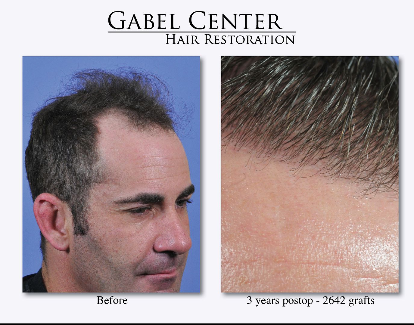
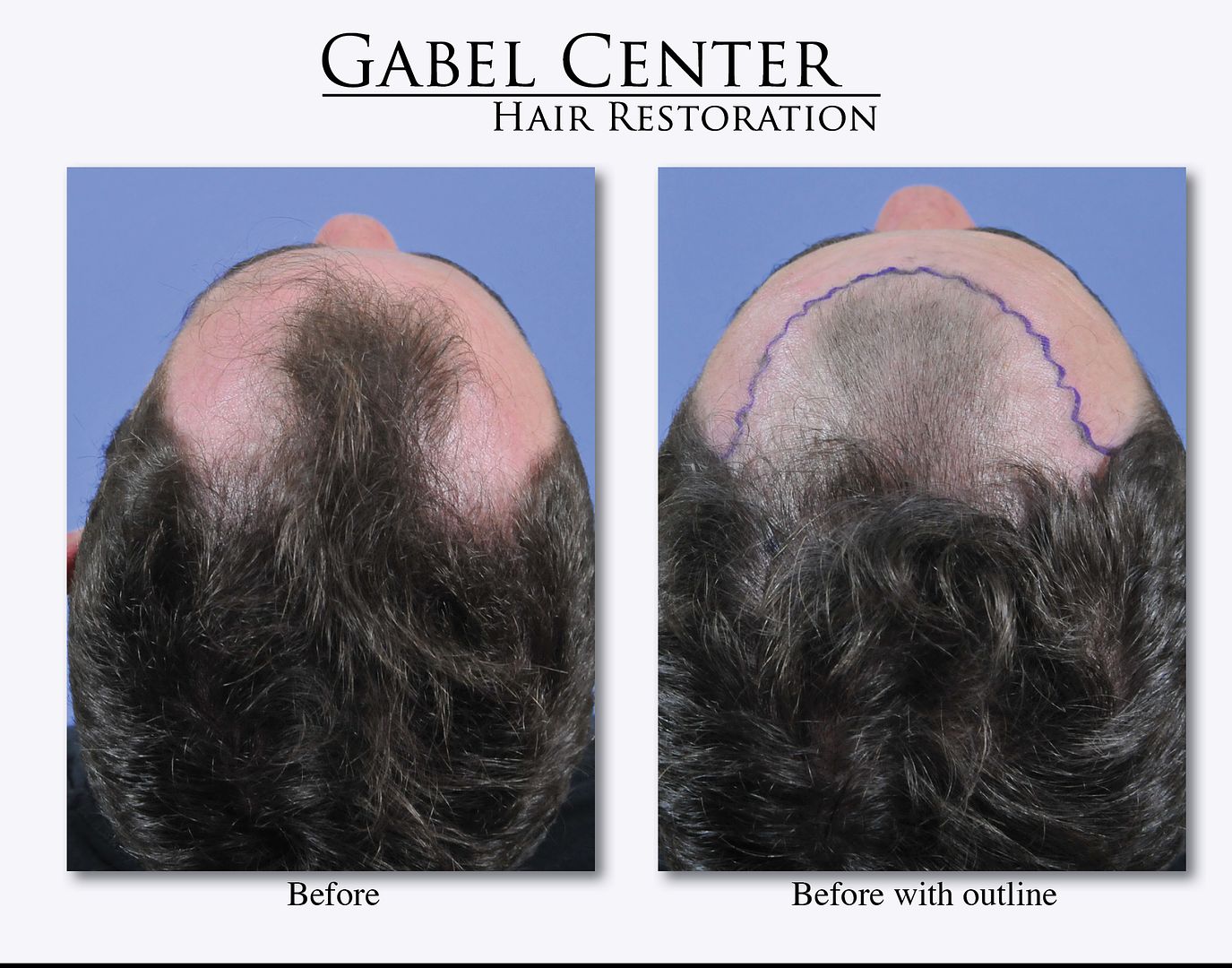
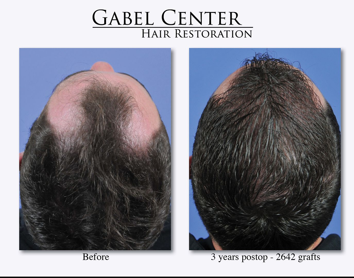
This patient is a 42 year old who is now 3 years out from 2642 grafts to the frontal hairline region. After 3 years, he is holding onto his native hairs pretty well as he is on finasteride and minoxidil. He styles his hair as shown with a spike even with short hair. The gel that he uses also gives a "wet" appearance which usually makes the hair look thinner; despite that, his hair still appears thick. At this point, he has no plans for a future transplant as this accomplished his goals and medical therapy is maintaining his native hair very well.
The photographs show our standard views with several very close up shots (the camera was only a couple inches away with a macro lens) illustrating the dense packing of the frontal hairline and the micro-irregularity pattern used to create the natural hairline with this patient.
-
Your planning shows with the great result.
-
Dr. Carman
Great result. There is always some uncertainty when grafting into scar tissue. The fact that you were able to do high density grafting into a scar with this result is excellent. Really nice job. Did you to a few test grafts first to see if they would grow prior to implanting the full scar?
Steve Gabel, MD
-
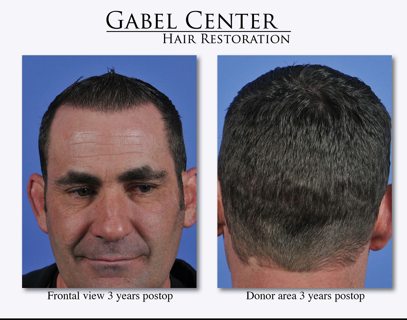
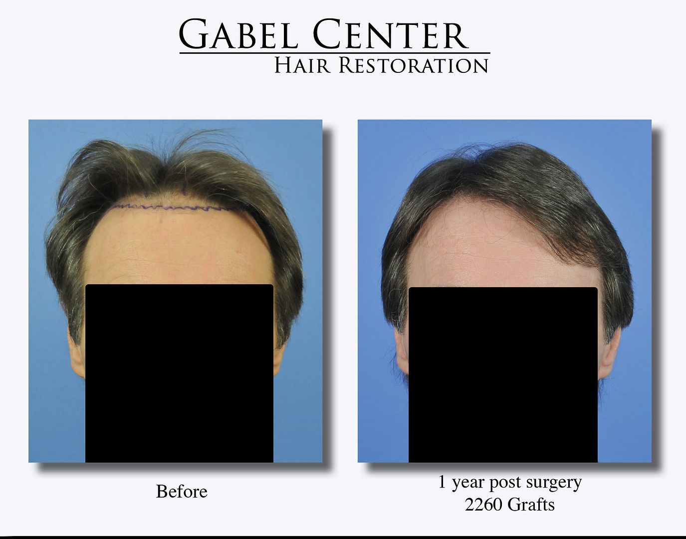
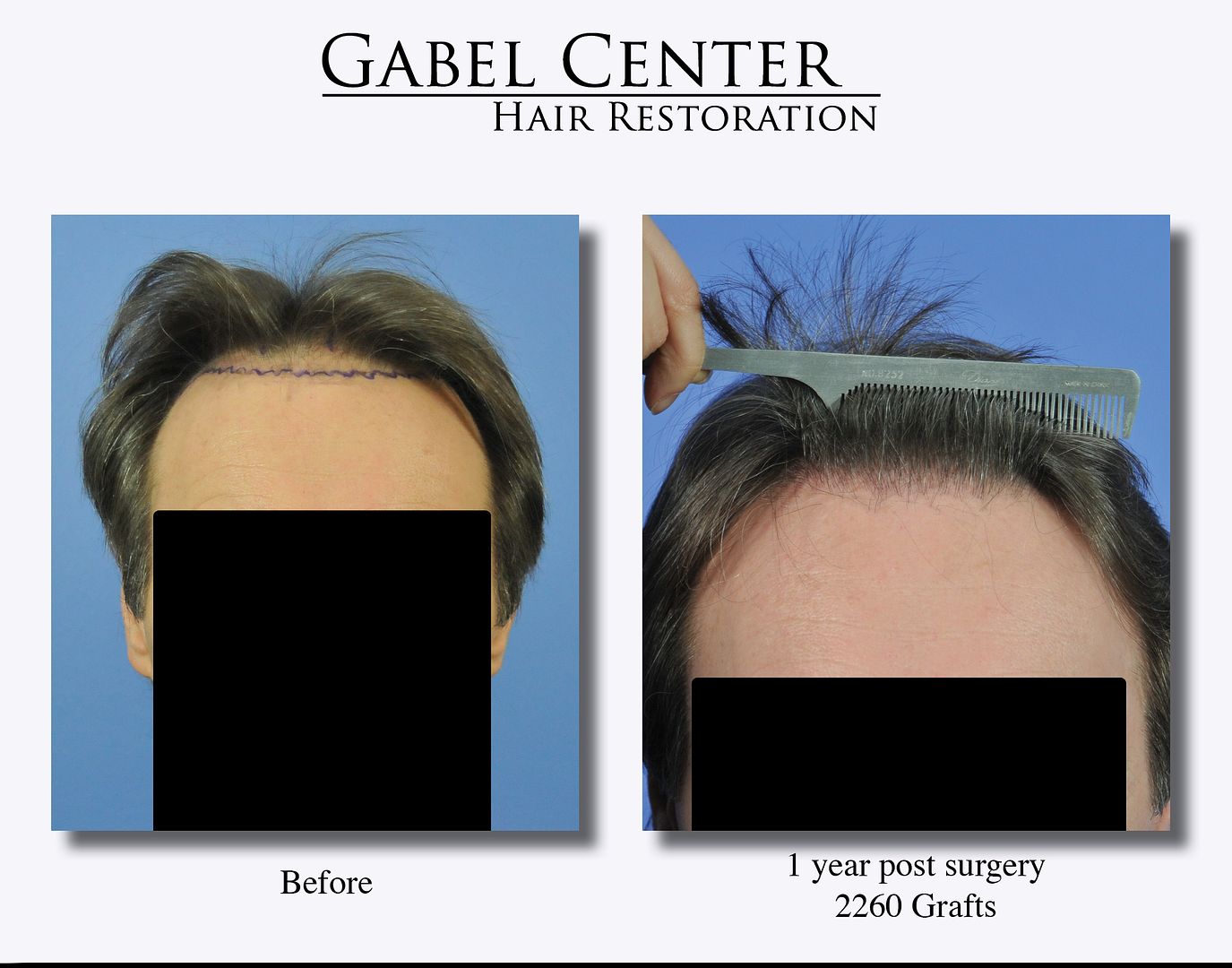
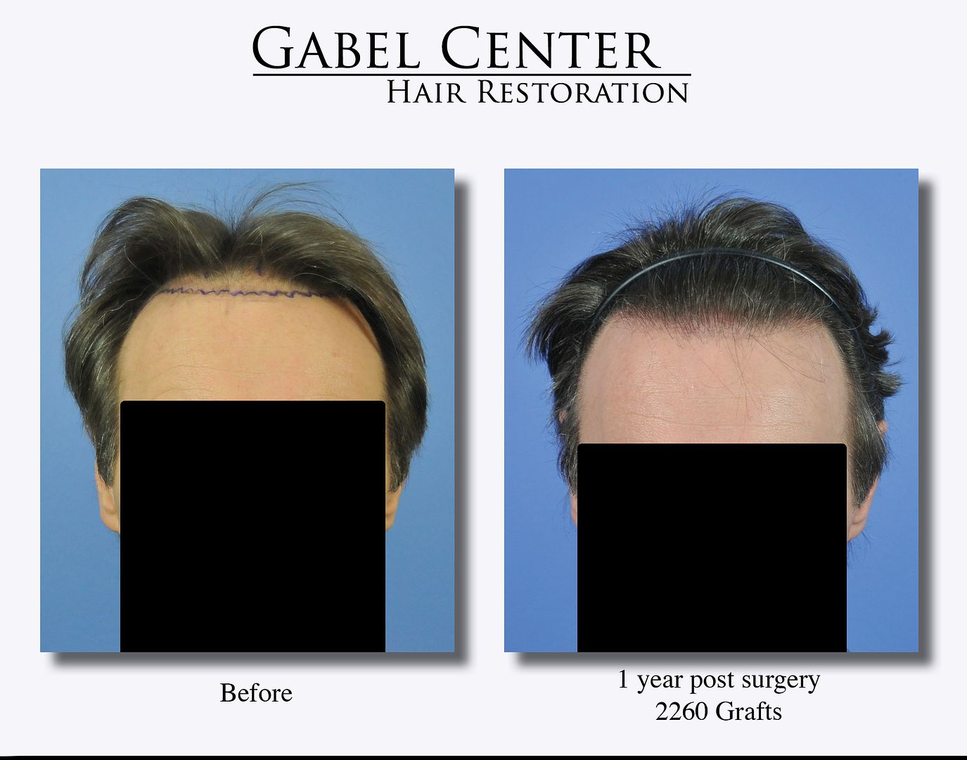
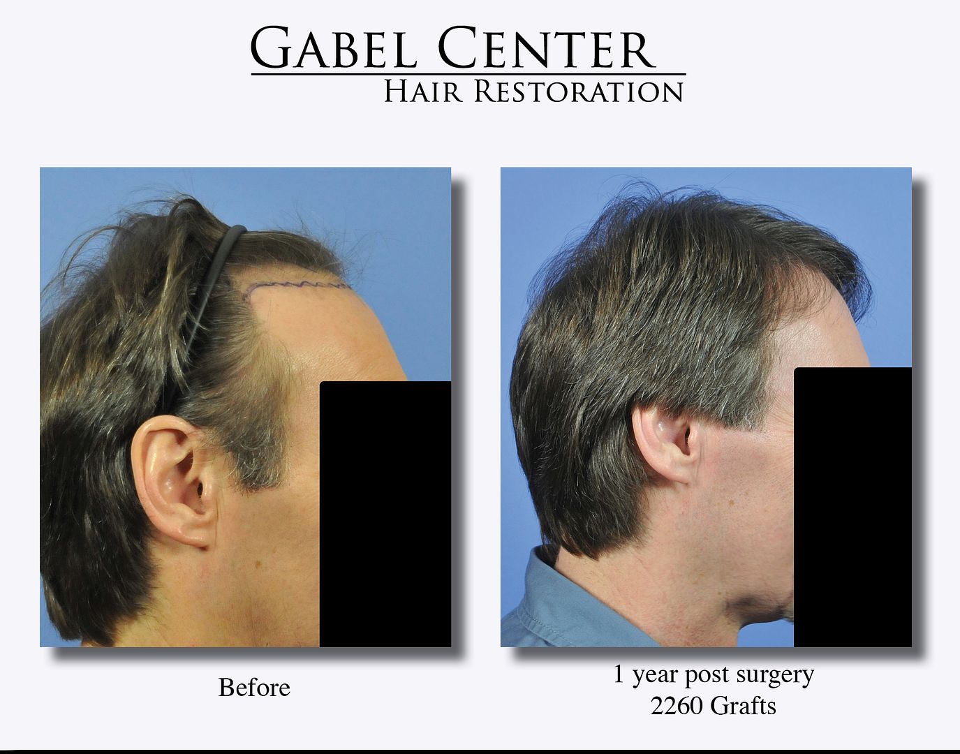
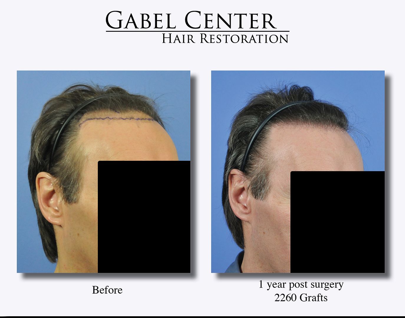
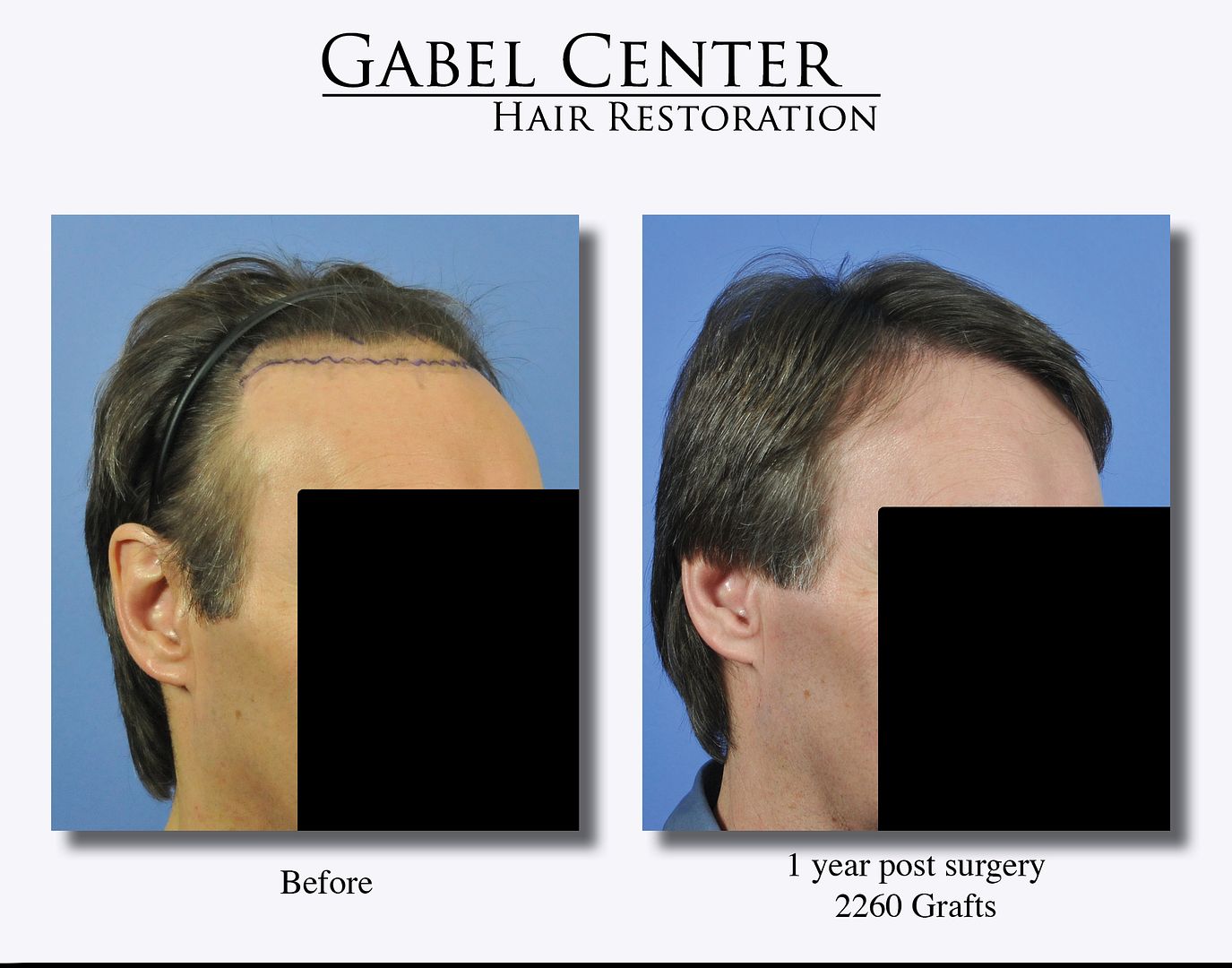
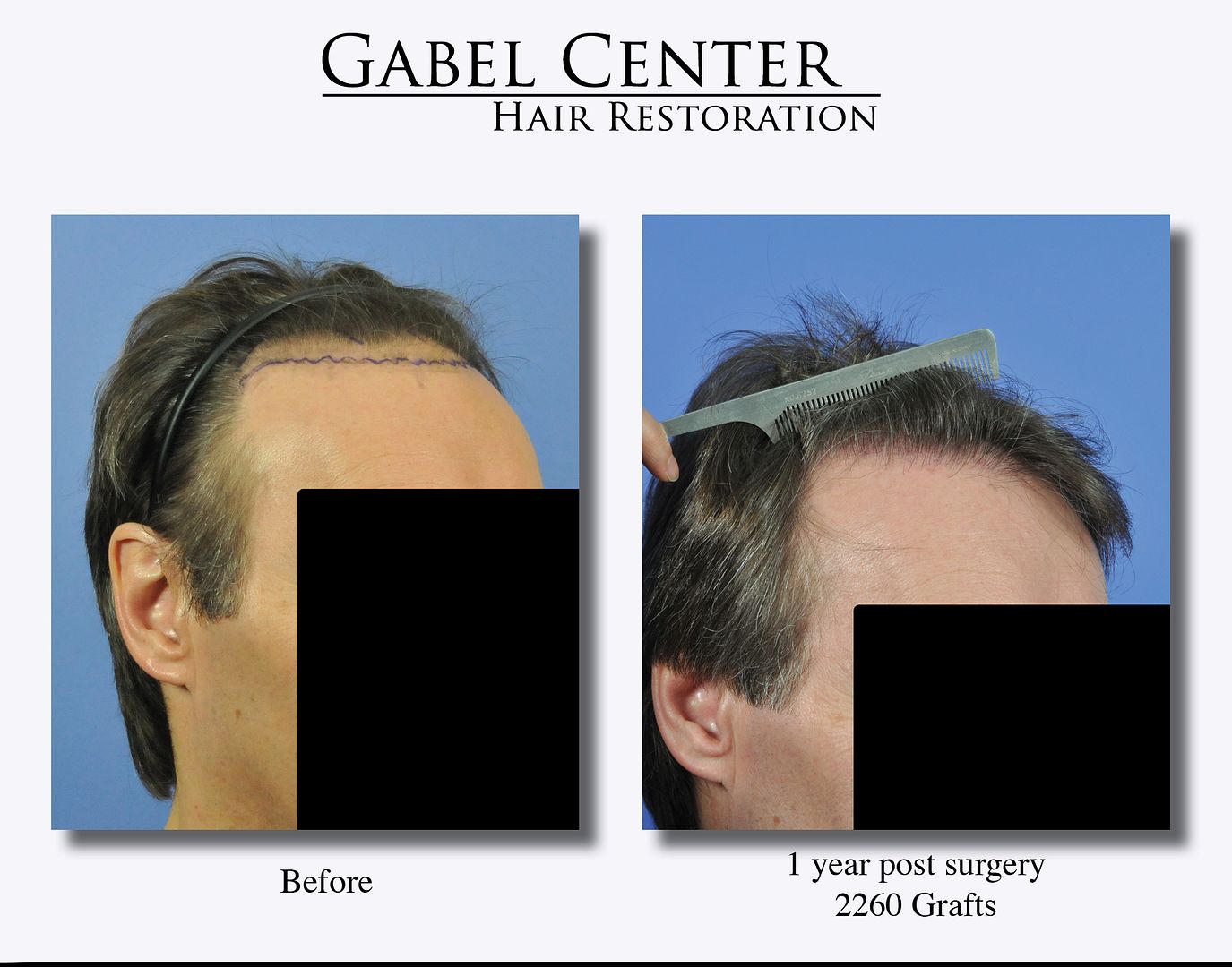
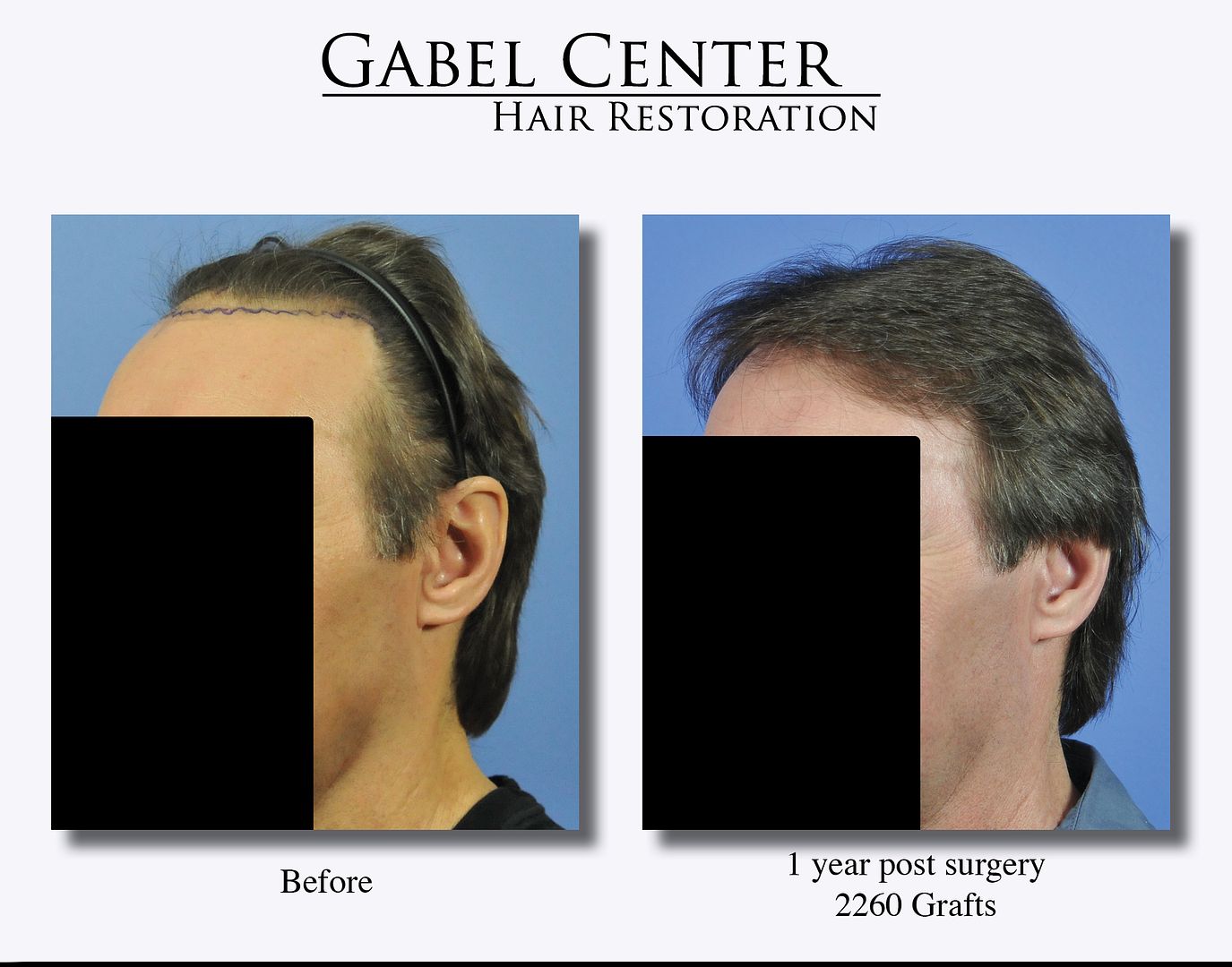
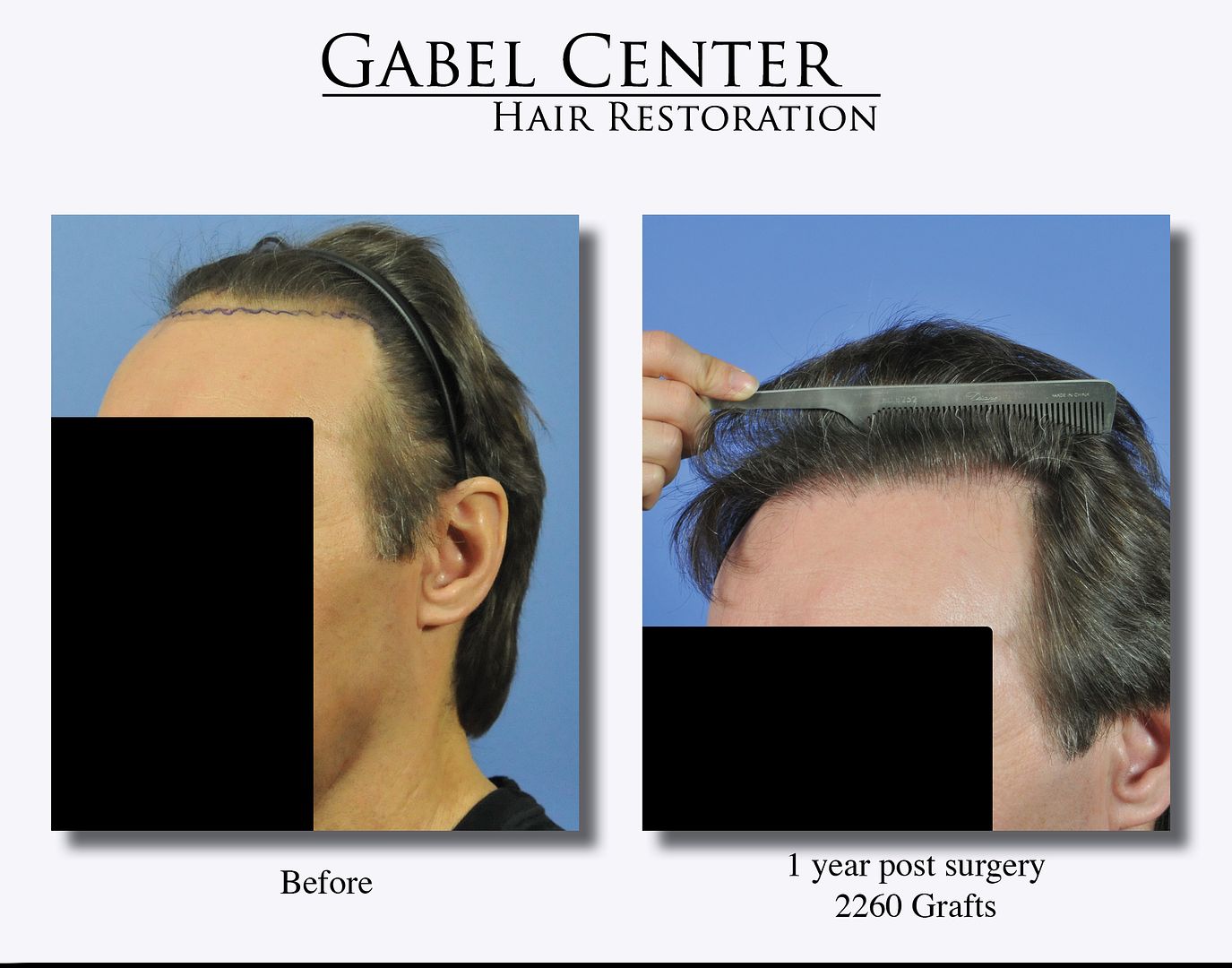
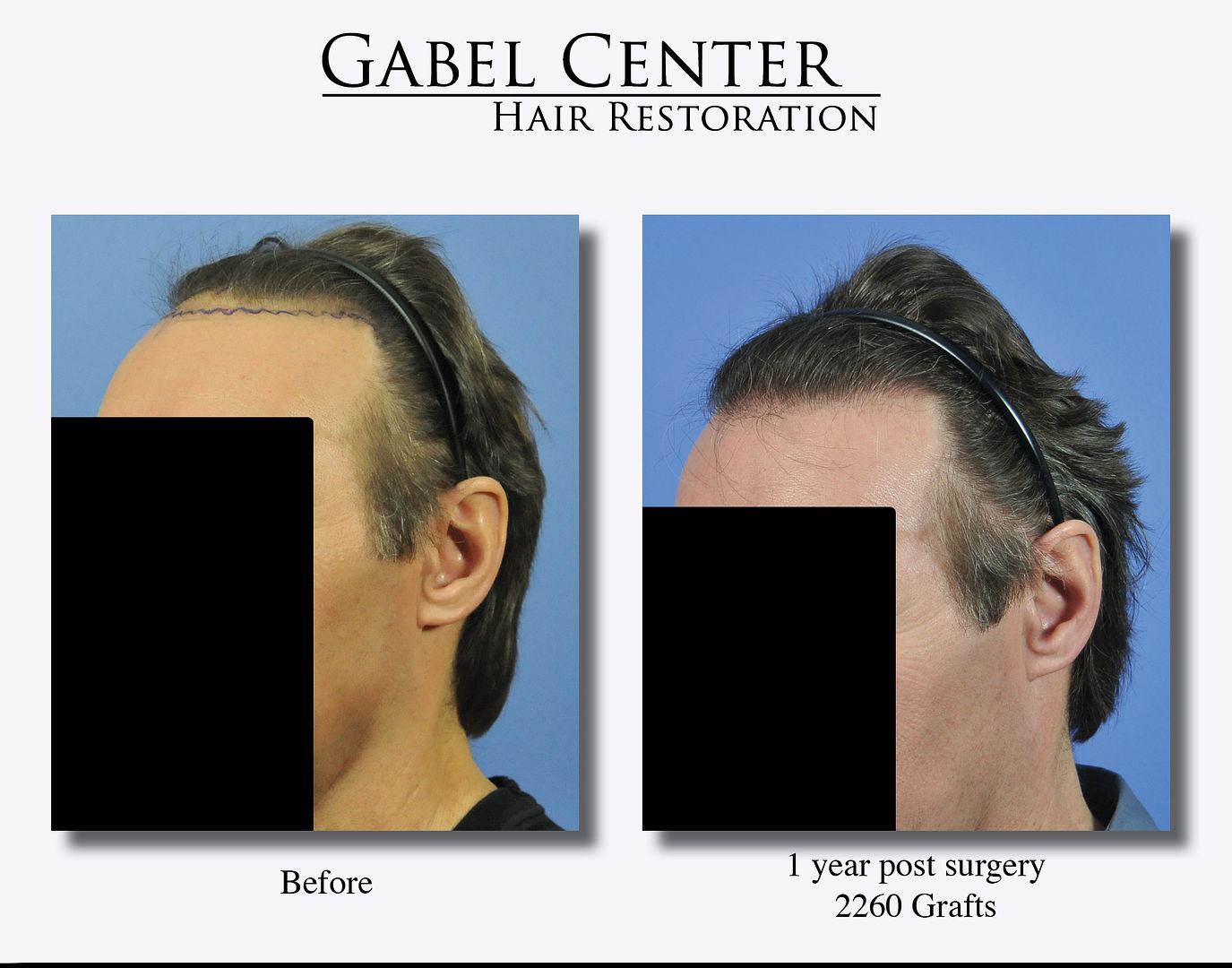
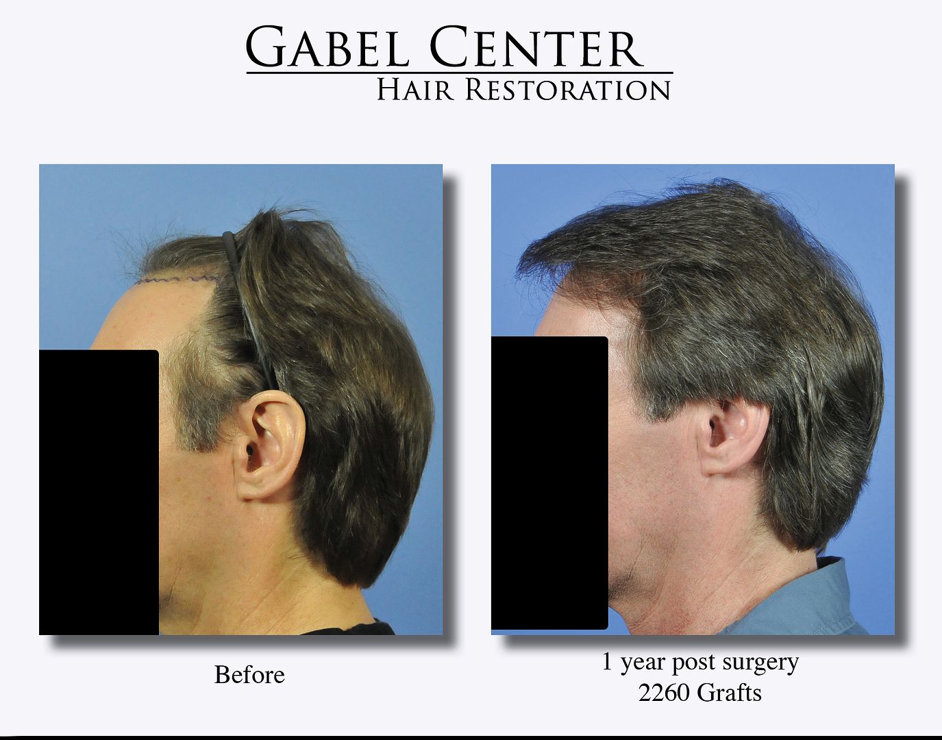
This patient is a 51-year-old male who desired to strengthen his frontal hairline. He has very good density in the frontal central tuft area of the scalp, mid-scalp, and crown. 2260 grafts were placed in the frontal hairline region.
The postoperative photographs show the 1-year result. Multiple photographs show the hair as he normally has it, and several depicting the hair with a band and comb to show the transplanted area more clearly.
Case details:
Total grafts: 2260
Strip method: 16.0 x 1.4 cm strip
Grafts:
1’s: 516
2’s: 1041
3’s: 568
4’s: 135
-
I appreciate all the positive comments. This patient is extremely happy with the results that he obtained after 1600 grafts. For the amount of hair loss and the area, I could have transplanted 3000 grafts to obtain even better coverage with strip; however, he desired to wear his hair short at times and therefore we performed FUE on him. His hair caliber was average to slightly thinner, which for the frontal hairline and thin appearance, is to his advantage so there is not a mismatch between degrees of alopecia and the hair caliber.
StaggerLee123: I agree – the shingling effect helped this patient.
SADbutTRUE: This patient recently returned so I wanted to show that I certainly do FUE. I’ll get some other cases posted as they grow out.
Future_HT_Doc, GBU, Cant decide, chrisdav: Thank you.
Spex and Mick: I appreciate your positive comments.
Plausibleboss: To answer your question, in the preoperative photo – he has very little hair to no hair in the frontal zone which is marked out with a purple marker, and in the post op photo, he has hair in the zone that was transplanted via the FUE method. Additionally, there is a close up photograph of the frontal hairline, which was transplanted, to demonstrate the growth and result.
Spanker: Thank you for your positive comments: it looks completely natural which is what we all strive for with each case.
-
The video is now added to the post.
-
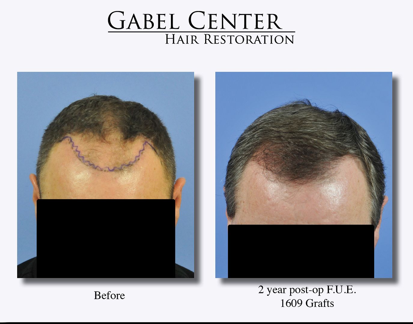
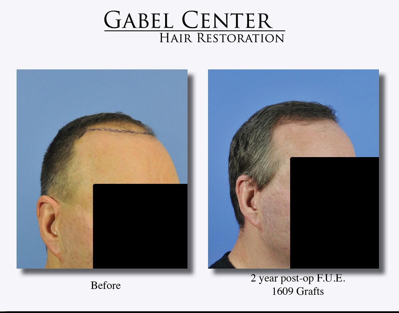
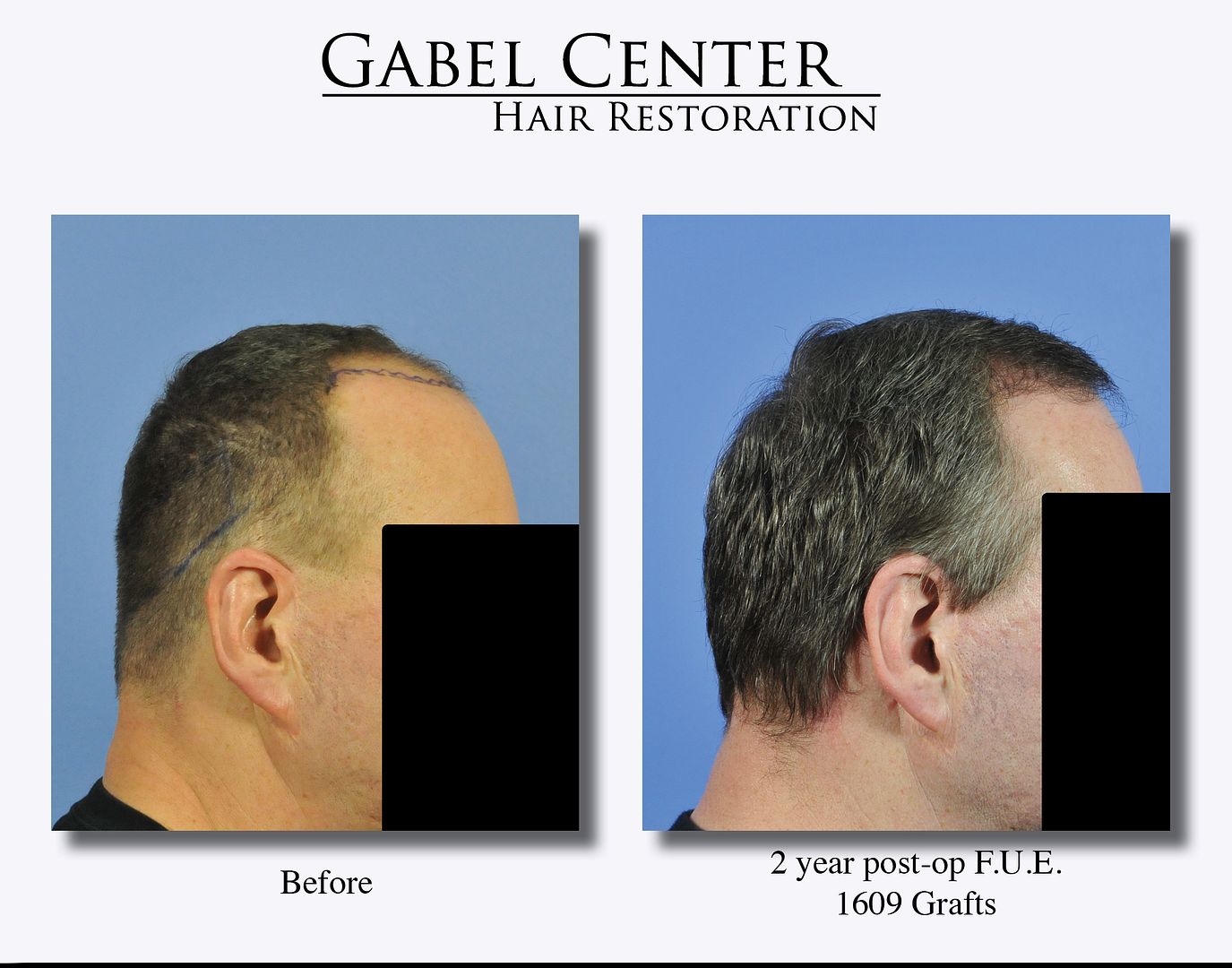
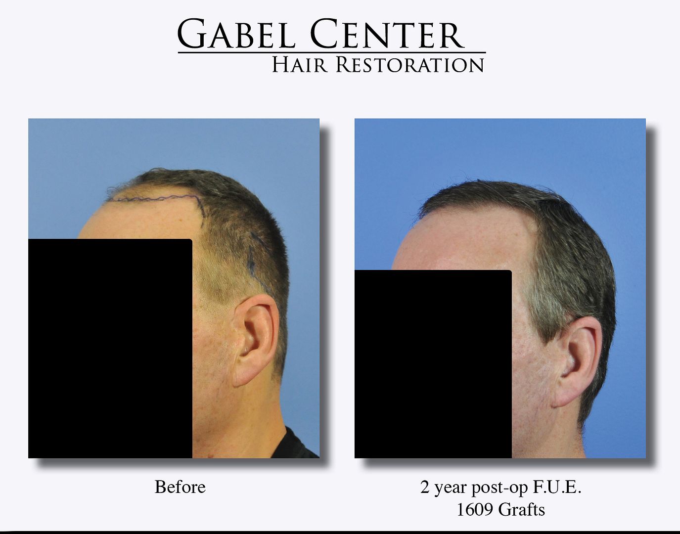
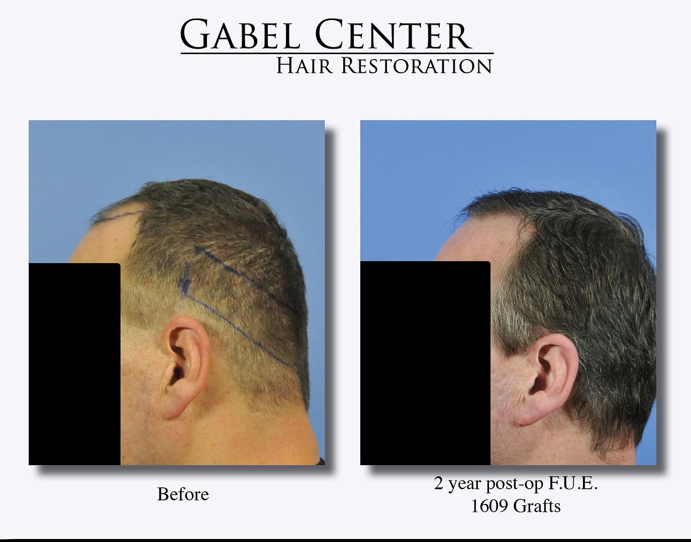
This 56-year-old patient two years ago desired to have his frontal hairline and forelock restored. His goals were very realistic in that he wanted to frame his face, and to have it appear natural. He wanted a hairline that was augmented, but age appropriate. He was also in situations when he had to shave the back of his head, and still is.
In discussing the options, I felt he was a perfect candidate for FUE. He understood that FUE is not a scarless procedure despite what is falsely advertised on the internet and TV, or what is read in highly deceptive airline magazine advertisements - it does leave hypopigmented areas at the graft extraction site, otherwise known as a scar; however, if the person wants to cut his hair down to only a few millimeters, then this is a viable option to cover the scars, but it does not eliminate them. I explain this in detail to every patient when discussing FUE. The preoperative photos show the proposed frontal hairline that he desired.
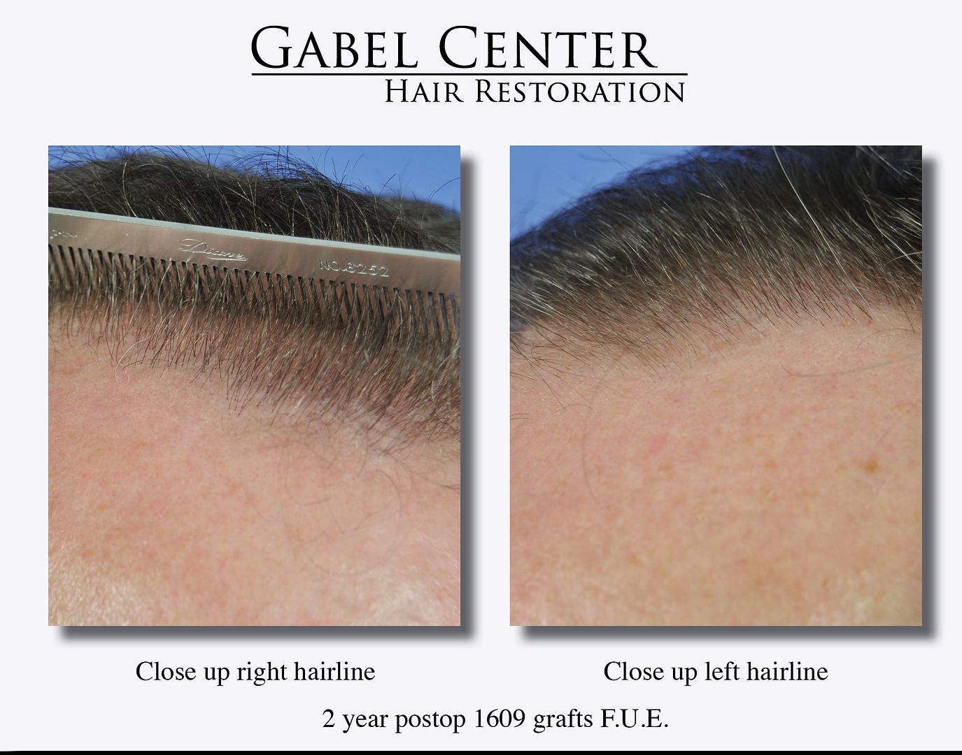
The photos show the results after 1609 grafts were placed into the frontal hairline and frontal scalp utilizing the FUE technique. I also have a video that I’m working on which will be added to this post in the near future which shows the extraction sites.
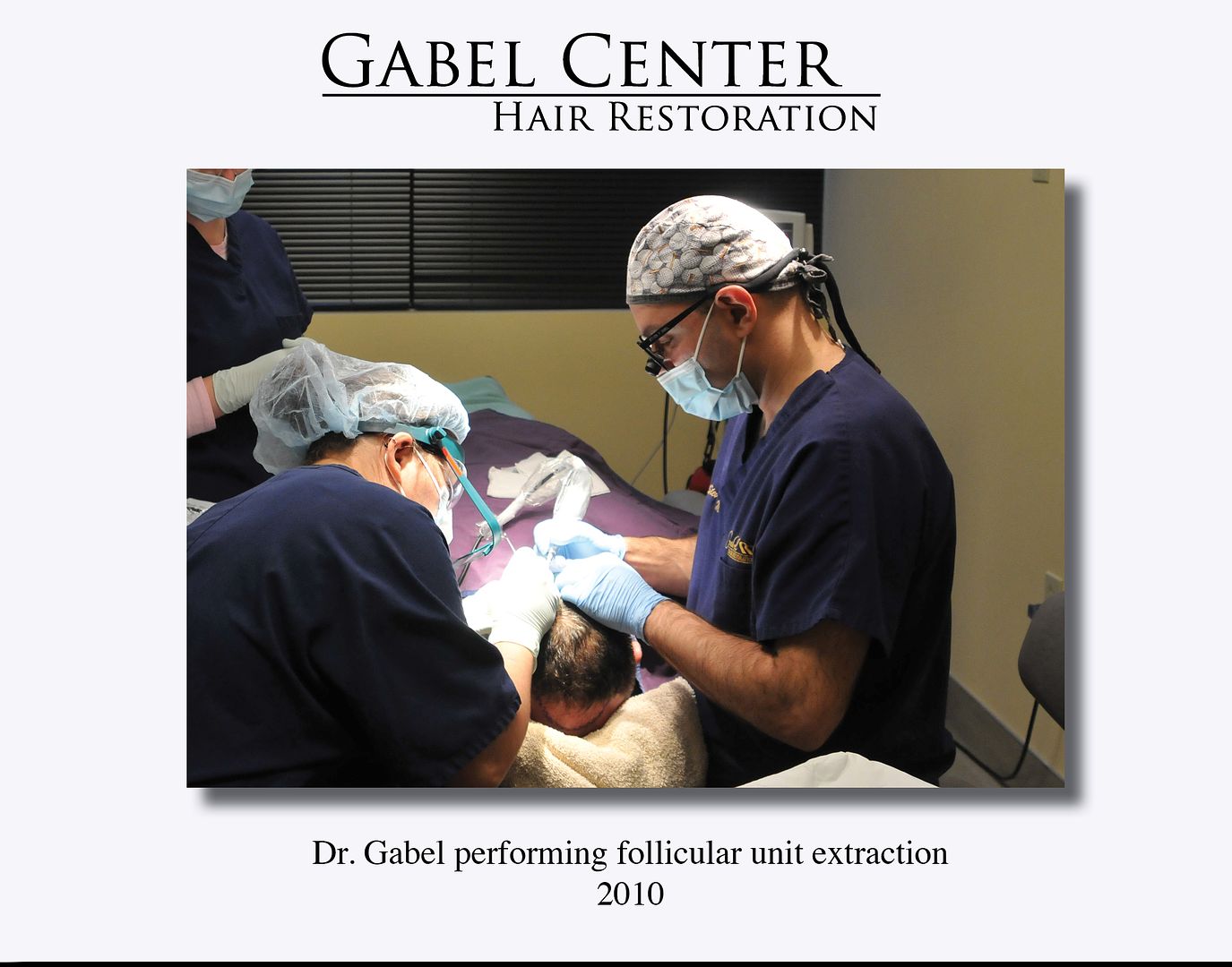
When performing FUE, I personally do all the FUE extractions on my patients. Since 2011, I use much stronger loop magnification to visualize the follicles for a more exact extraction process. Once the grafts are extracted, they are carefully examined under the microscope for any signs transection or visualized damage to the grafts. If any damage has occurred, they are rejected. This patient is extremely happy with the results as it accomplished his goals.
-
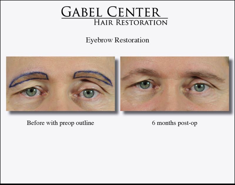
This patient was interested in an eyebrow transplant to increase the density of his existing sparce eyebrows. He stated that he never had very thick eyebrows so his goal was to define the eyebrows more especially in the outer half of each one. He did not want them too thick.
Approximately 230 single haired grafts were densely placed in each eyebrow. The hair direction is extremely important in eyebrows as the hairs must lay flat against the skin and in the correct orientation in specific portions of the eyebrow. The middle portion of each eyebrow is oriented up and laterally. The middle portion is oriented laterally, and the outer portion of each eyebrow is oriented laterally and inferiorly. If the hairs are not oriented properly, then the eyebrow does not look natural.
The before photos are presented with the outline desired by the patient. The post op photographs are 6 months out. As seen, they are growing well and will mature over the next 18 months.
-
SadbutTrue
Thanks for your comments. To answer your question about survival, I cannot give you an exact answer as that may be virtually impossible based on the number of grafts transplanted; however, the graft survival is very high based on my examination that there are not any areas of poor growth or density at this point. If my examination would show areas of poor/no growth, one may conclude that a patient had graft failure; but as you can see objectively in the photographs and the video documentation combing through his hair, the growth so far is going very well in all areas. Also, keep in mind, that he is only 6 months out from his procedure, so if there are any sparse areas, he is still in the early stages of hair growth and a re-examination in 6 months will give a much better representation if he did have areas that did not grow. So far with this patient, we are on track for an excellent result.
Overall, your question is spot on: it is so important to constantly evaluate patient results by objectively analyzing the progress of patient's growth. I take detailed notes on every procedure and when I see excellent growth like this patient, I review what I did and make sure I continue that technique. For analysis, I standardize my photographs as much as possible by using the same camera and lighting (I do not use flash) and taking each photograph so each view of the patient is as close to the previous photo taken by that same patient with a standardized background. In my room I have dedicated to photographs and video, I actually have markers in exact locations and angles where the patient looks so that the photographs are standardized and one can make an accurate evaluation of growth. When I can, I also take close up photographs with a 60 mm macro lens to see the fine detail of the frontal hairline, which is also demonstrated in this patient.
In terms of scars, I also try to take photographs of the donor scar, whether it is from a strip procedure or FUE procedure, and evaluate the results. On average, my scars are very small as seen in this patient. Again, I pay close attention to detail with my surgical technique to minimize the scars on patients. There are so many factors that play into the development of scars, many intrinsic to the patient (how well do they heal) and extrinsic (surgical technique). Again, attention to detail is king.
-
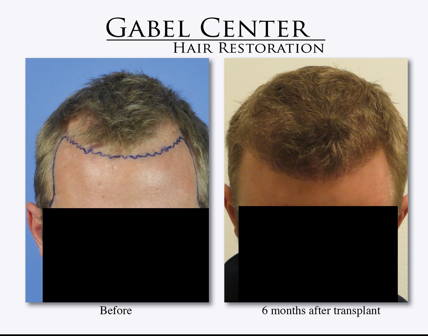
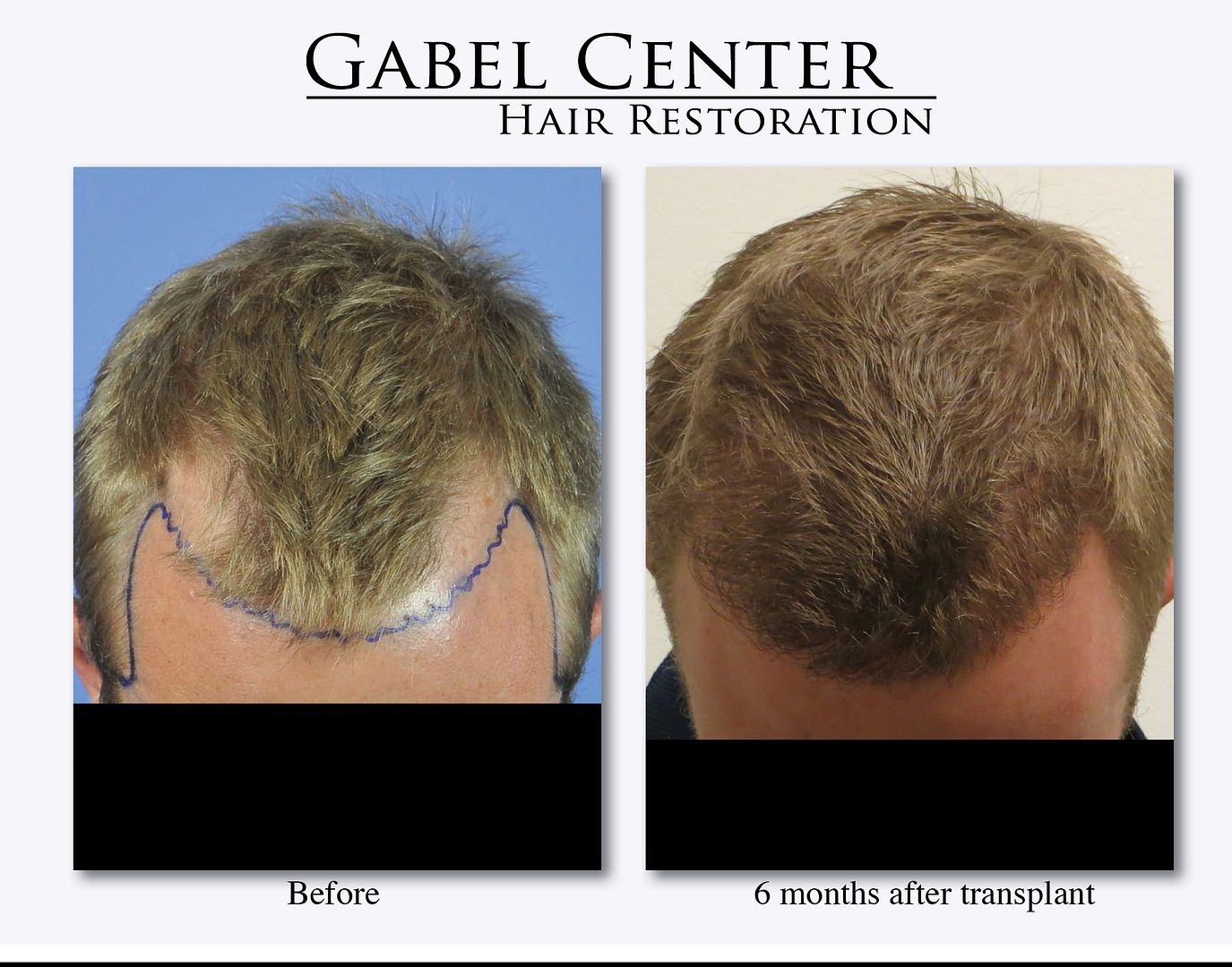
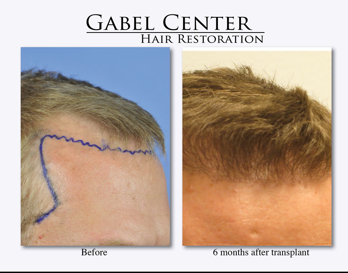
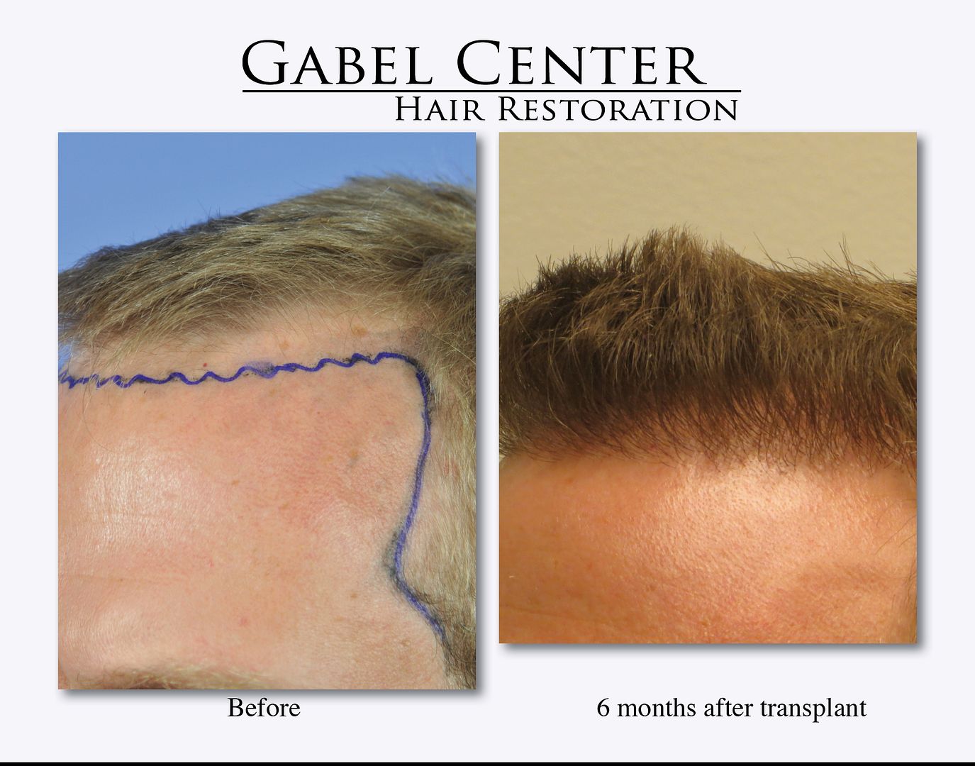
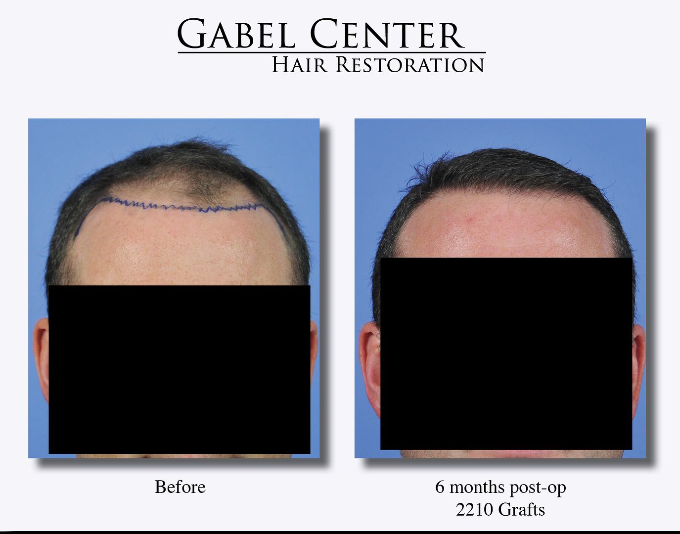
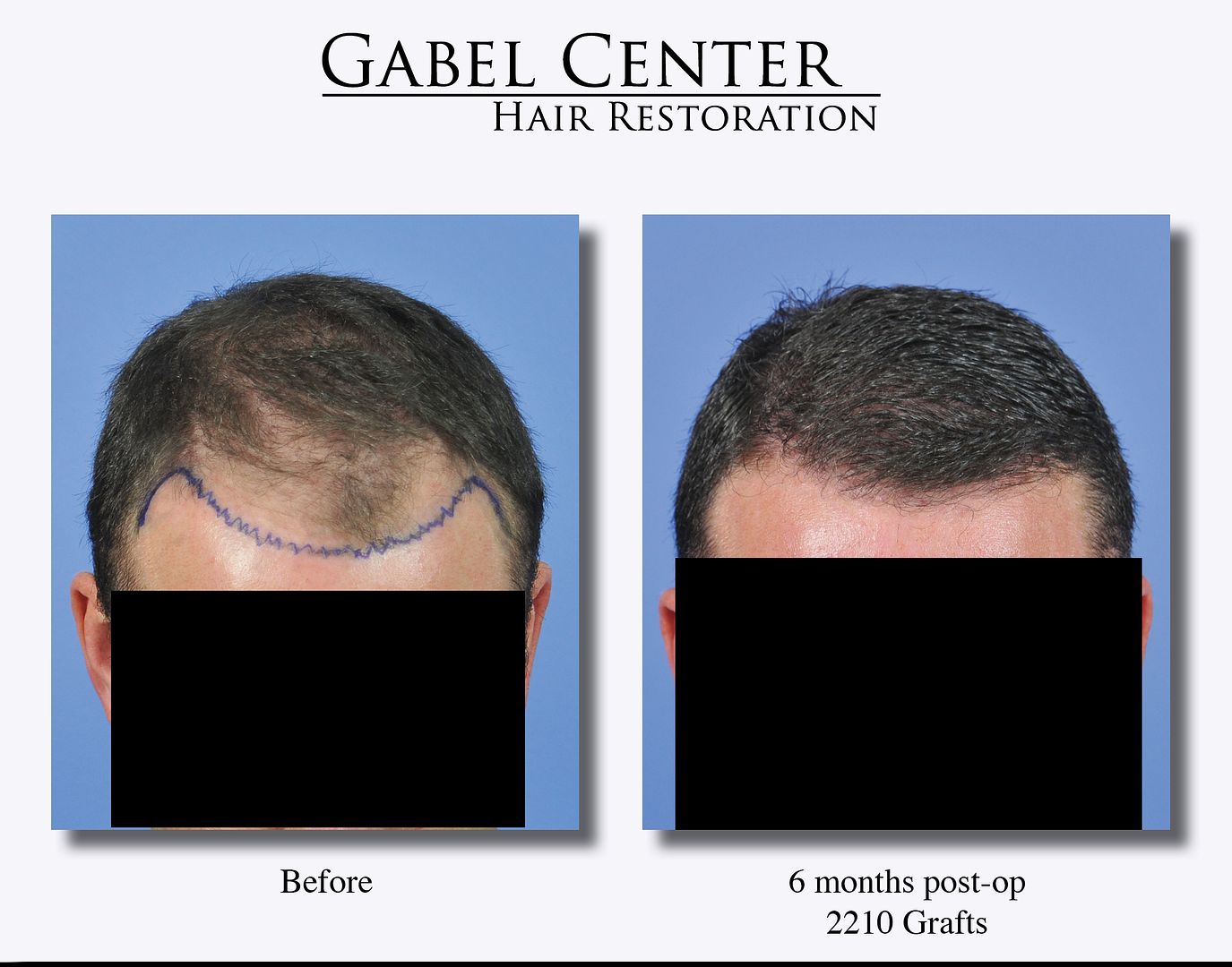
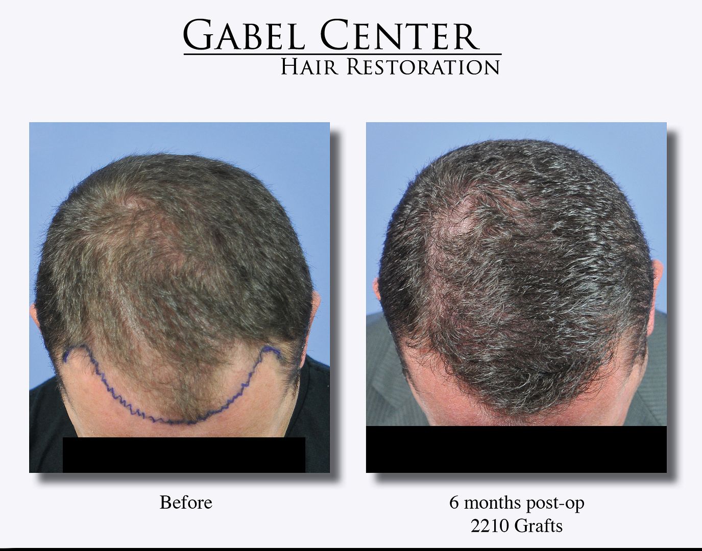
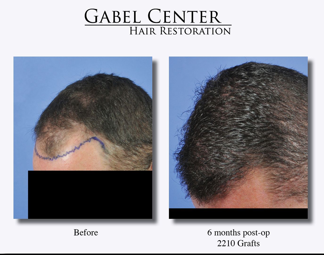
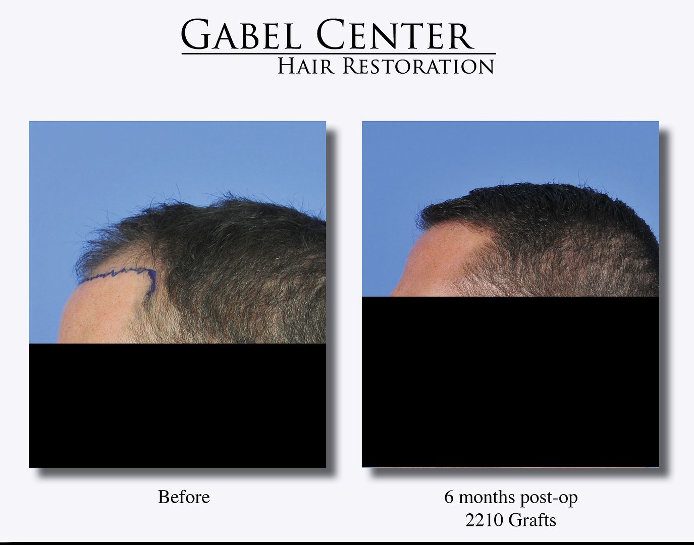
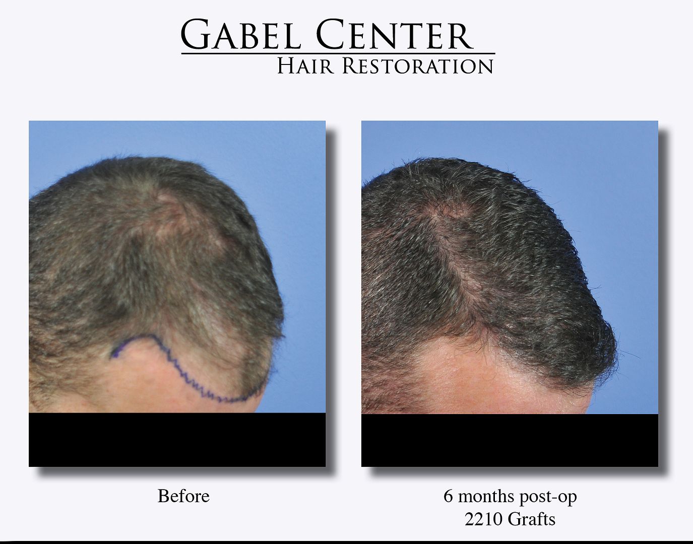
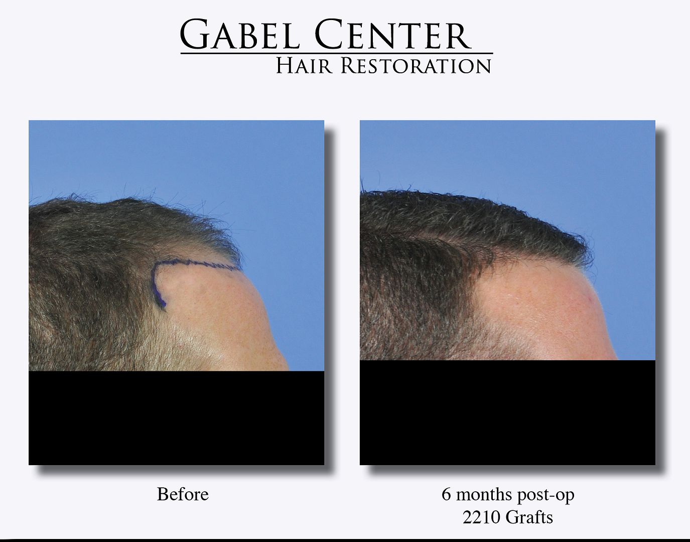
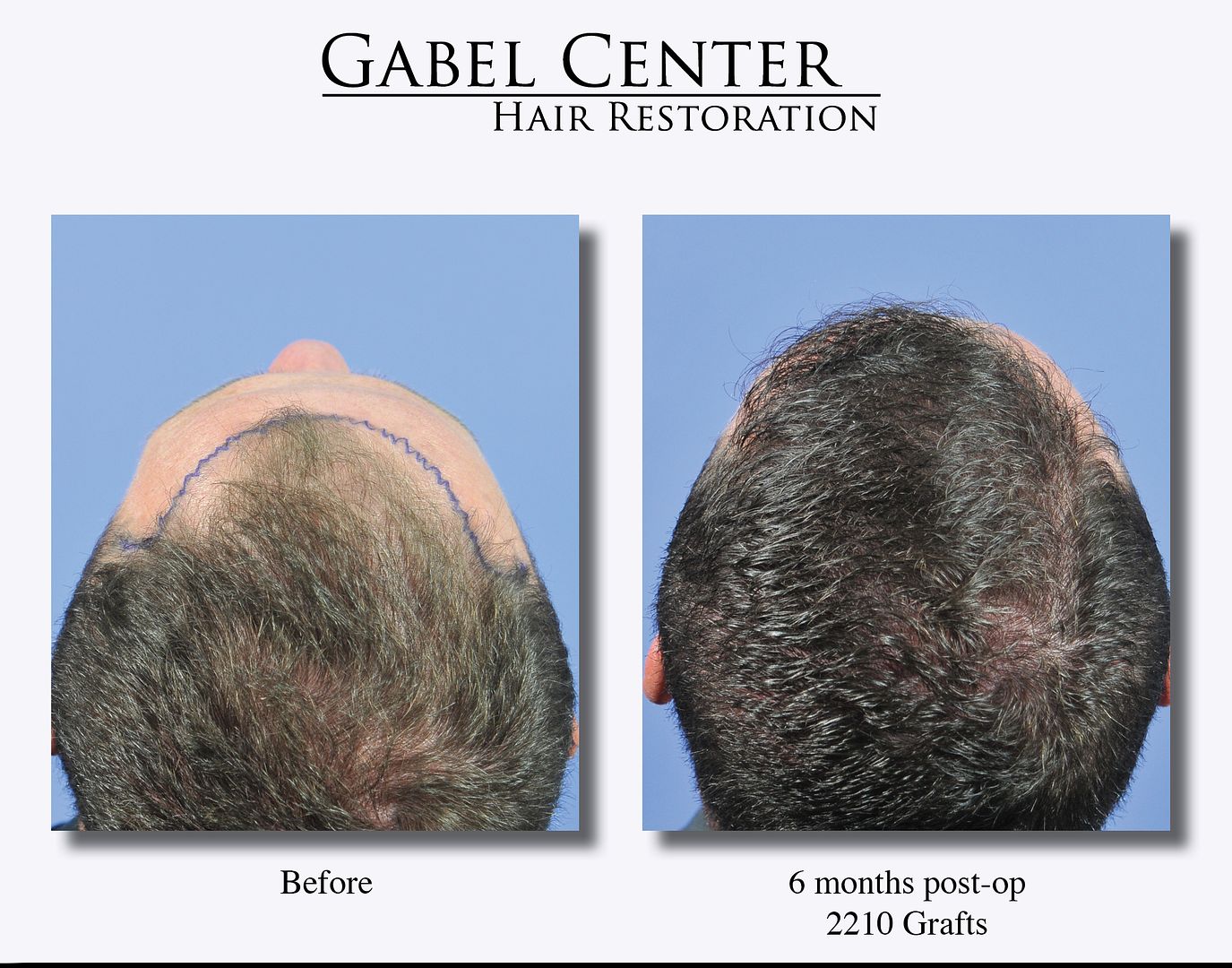
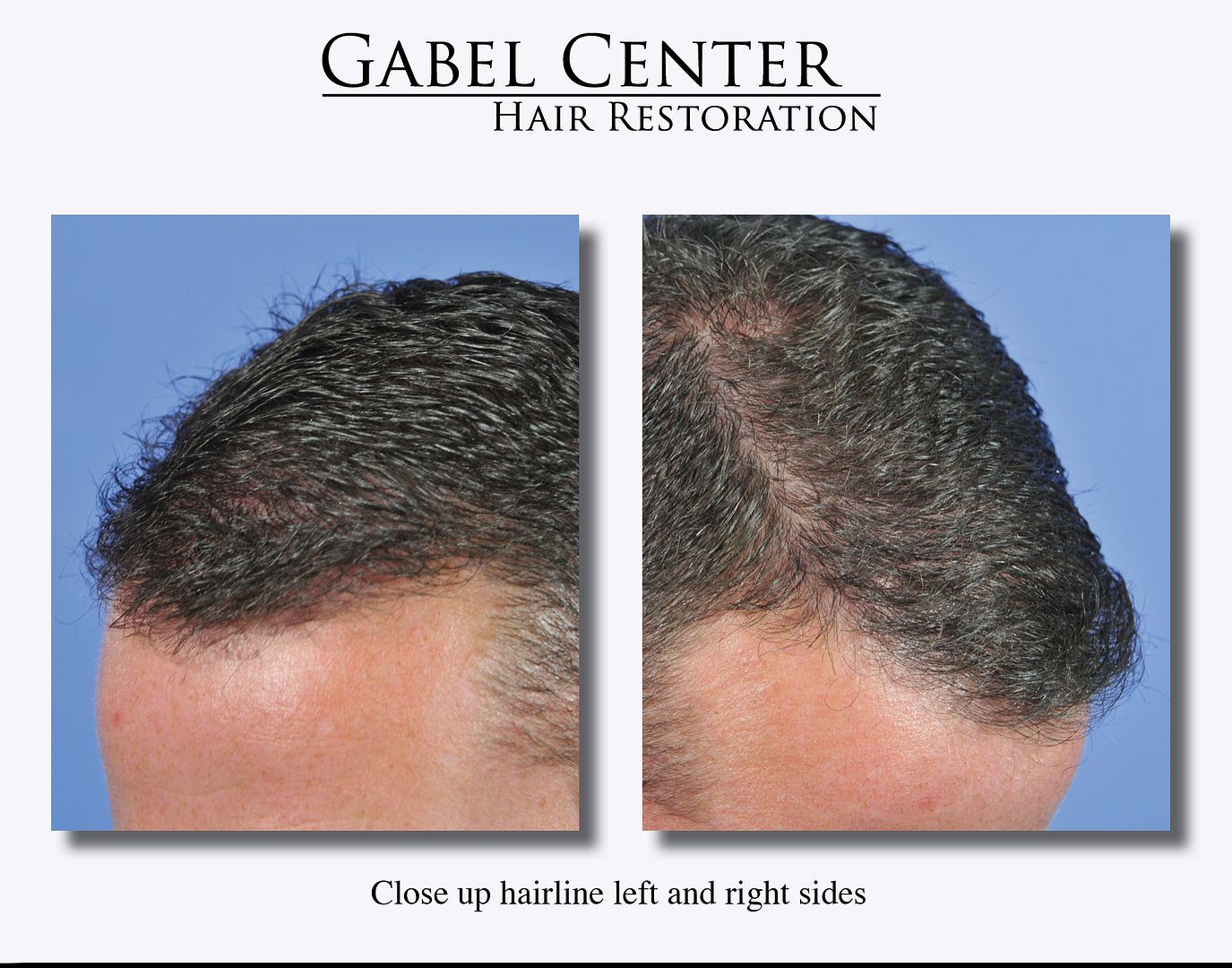
This 36-year-old patient desired to have his hair restored in the frontal region of the scalp and the frontotemporal angles. A total of 2210 grafts were placed into the frontal hairline, scalp, and frontotemporal angles.
Preoperative and postoperative photographs are presented at only 6 months growth. Close up photographs of the donor area show no evidence of the donor area scar despite the very short haircut. A video is also presented to show the results with a comb being run through his hair.
-
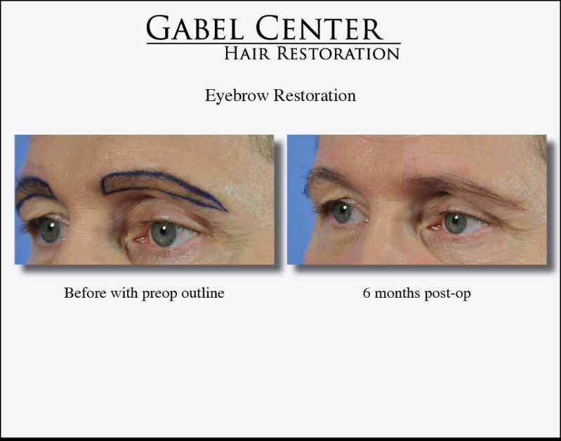
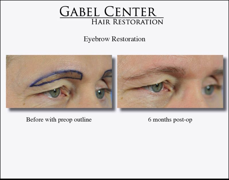
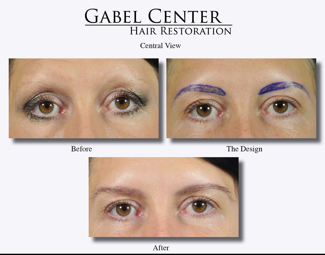
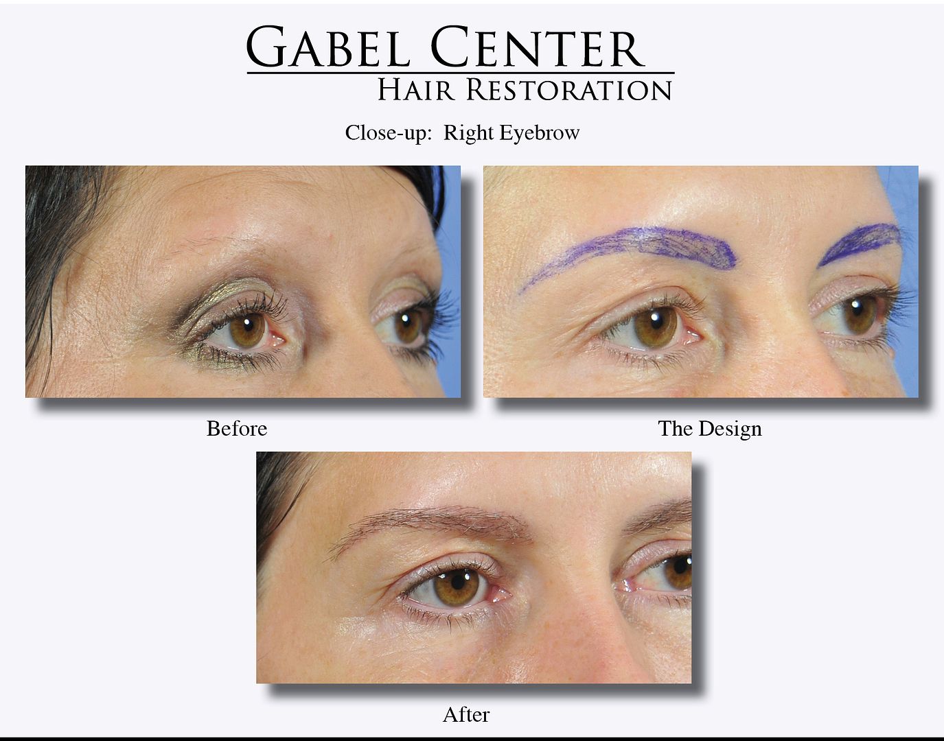
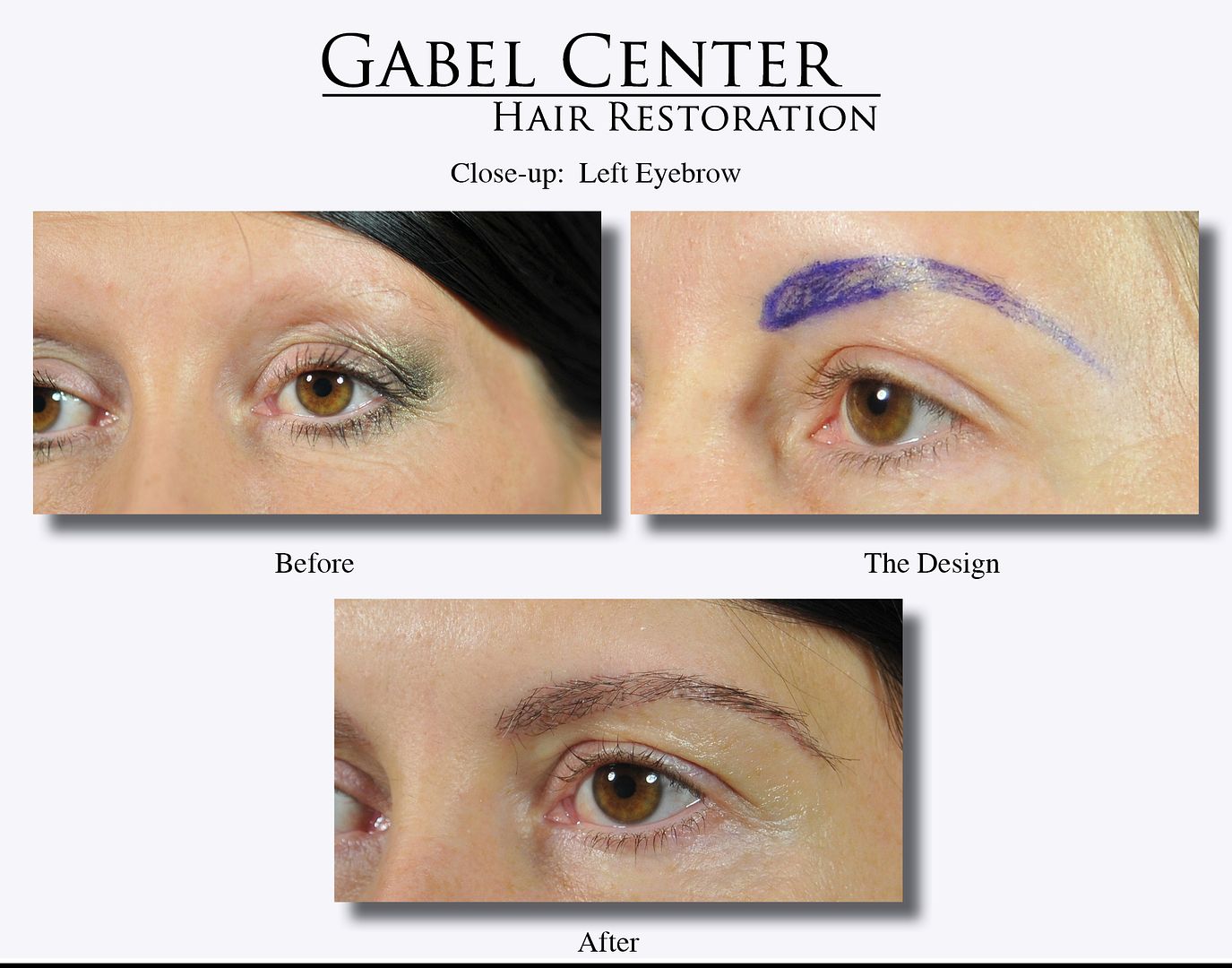
This female patient presented with only a few, scant hairs in each eyebrow. When she was a teenager, she thinned them out by plucking; unfortunately, after a few years of thinning them out too much, they never grew back in. Her goal was to restore her eyebrows so they provided a nice balance of her facial features and definition to her eyes.
To achieve her goals, about 250 single-haired grafts were transplanted into the eyebrow as shown in the design photos. I always have the patient draw their ideal pattern and adjust it for symmetry and naturalness.
From a technical point of view, it is extremely important that the hairs lay as flat as possible on the skin, and that the natural curvature of the hairs are oriented correctly or else the patient will have "wild hairs" that are coming out in the wrong direction. The medial portion of the eyebrows are oriented superior/laterally, and as the eyebrow sweeps away from the central area, the hairs are oriented laterally with the curvature pointed toward the central portion of the eyebrow. This type of design and hair placement produces a very natural appearing eyebrow as seen in the "After" photographs.
-
Thank you for the positive comments. This patient is extremely happy with her result; we do expect it to thicken even more in the next year.
-
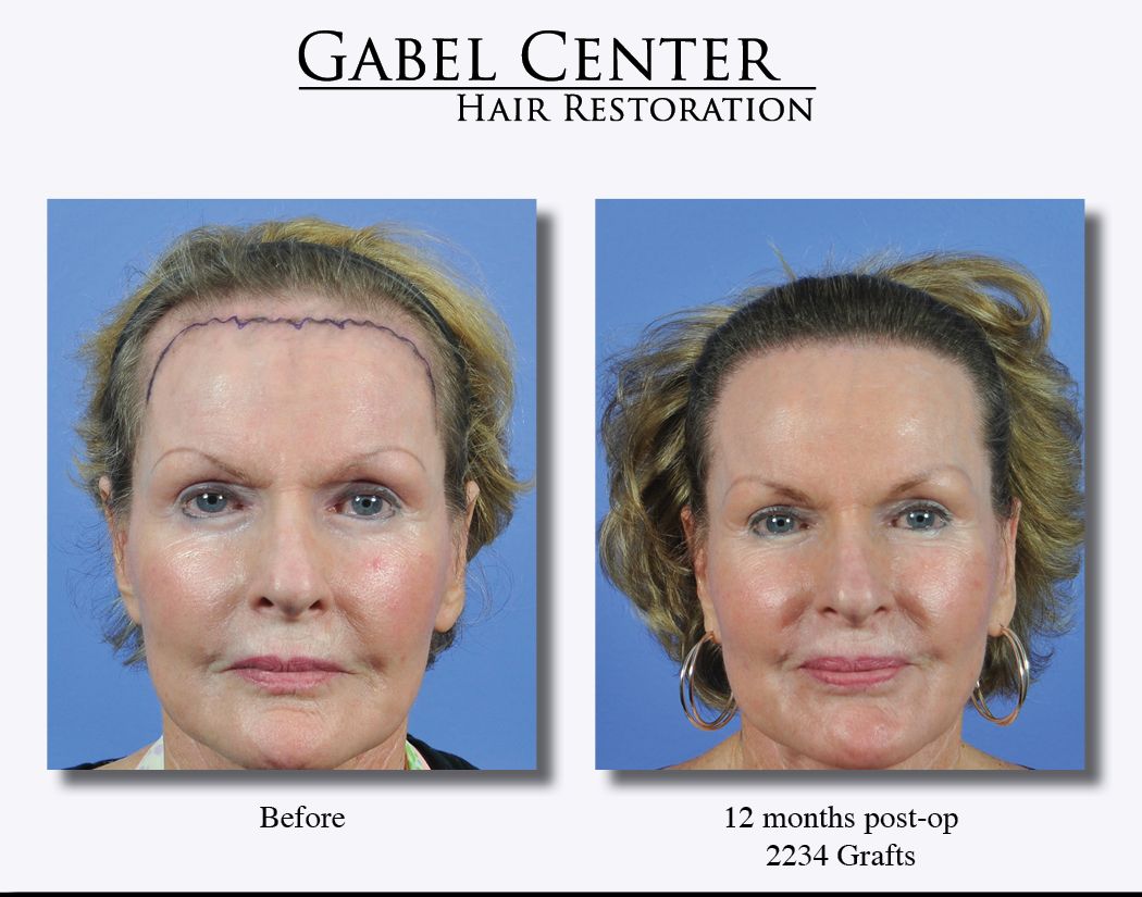
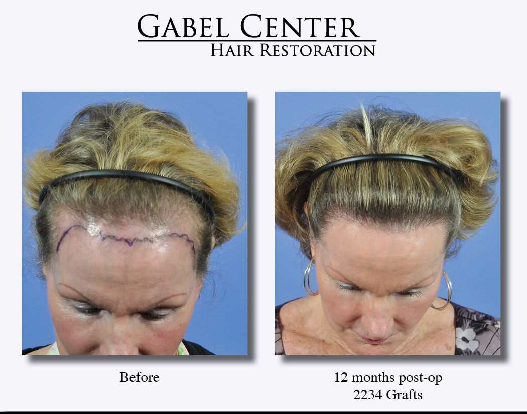
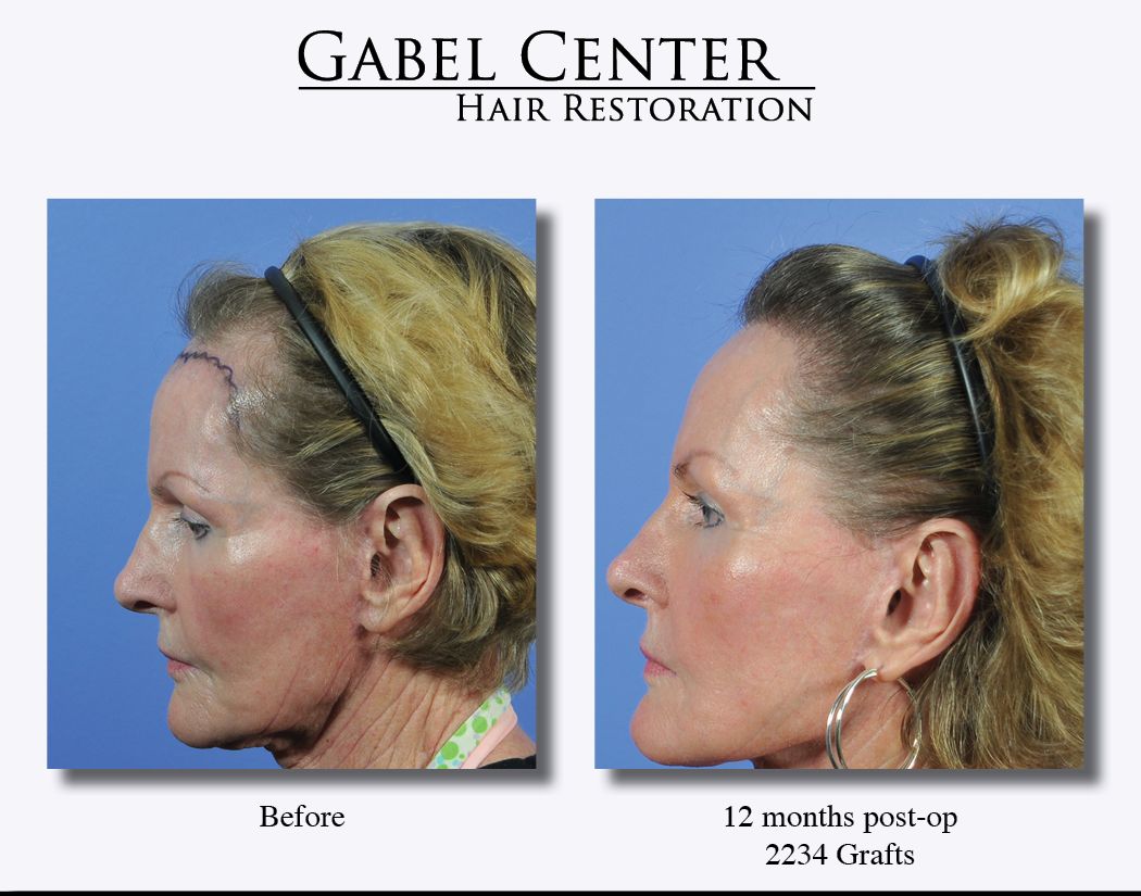
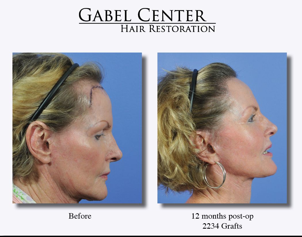
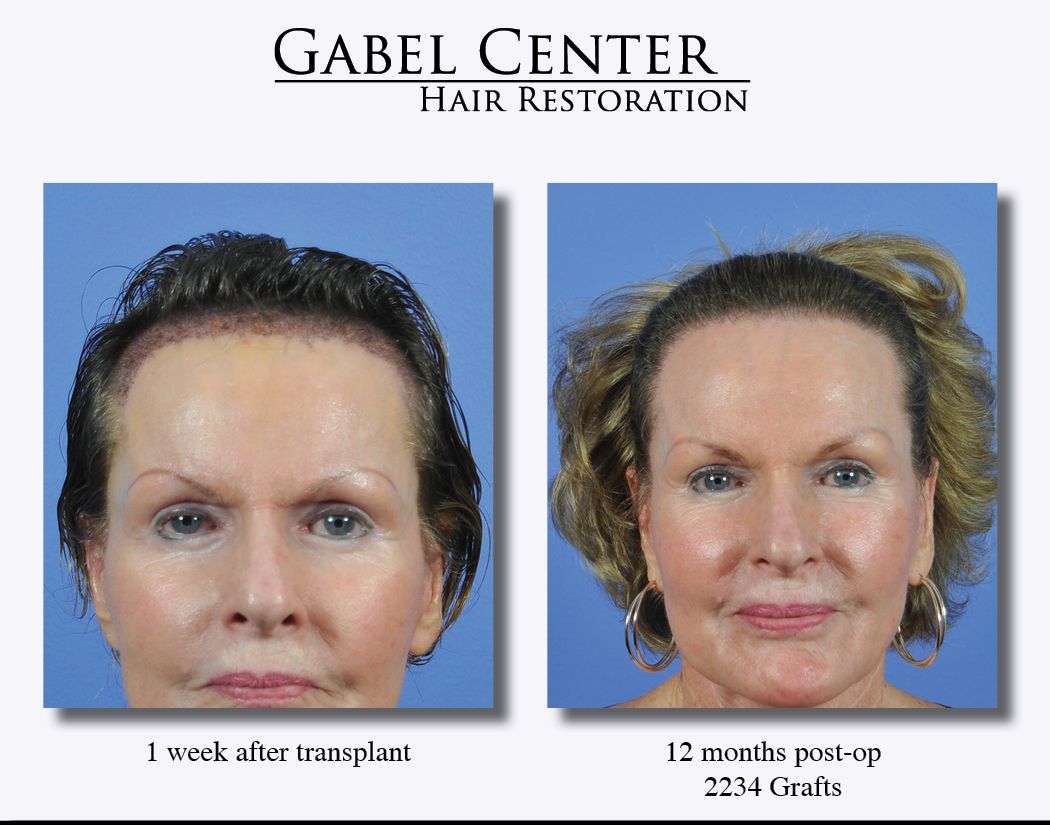
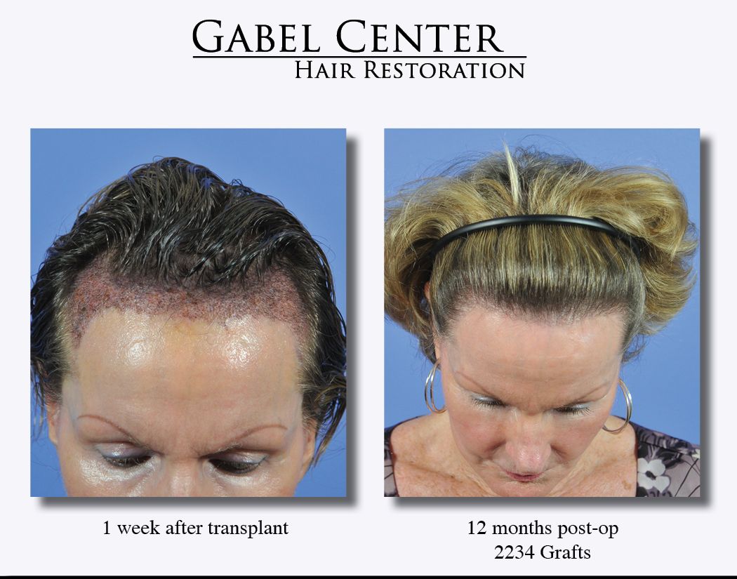
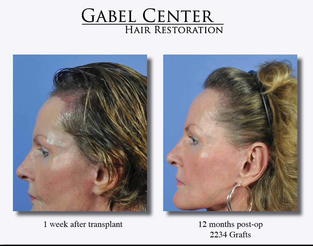
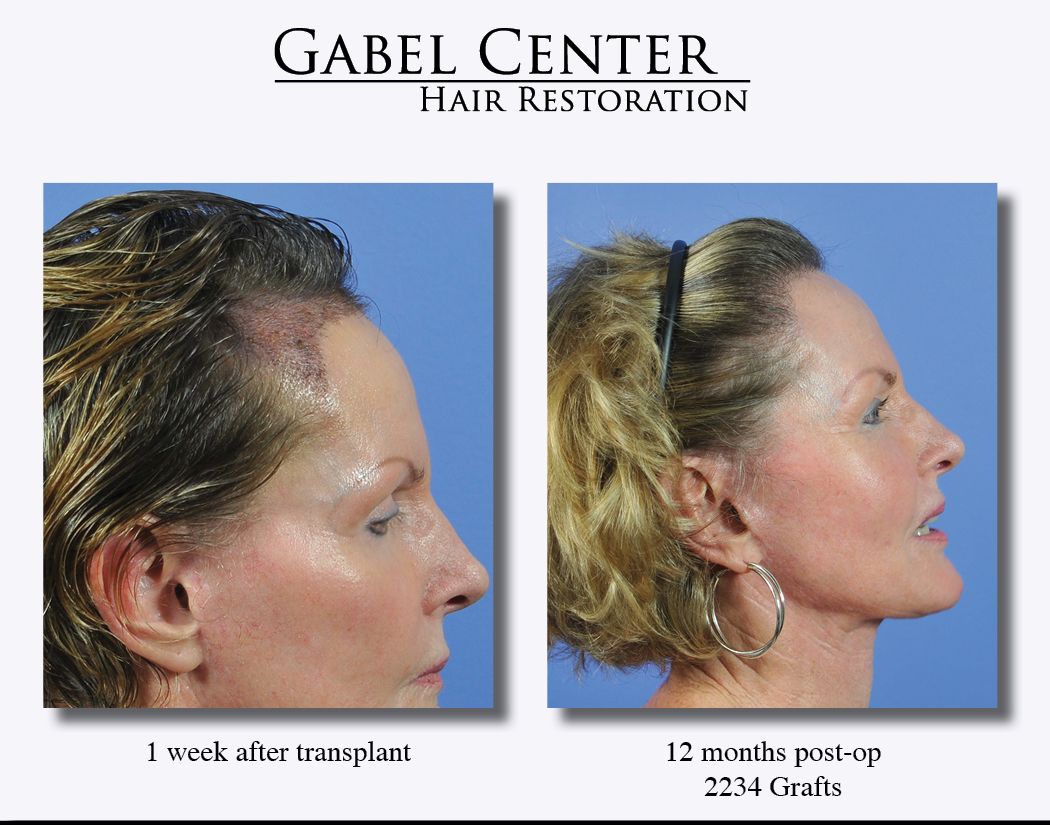
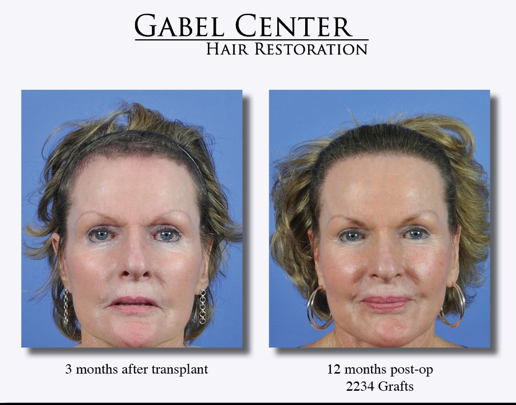
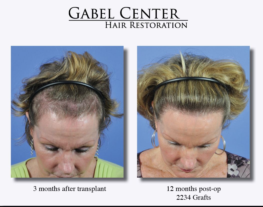
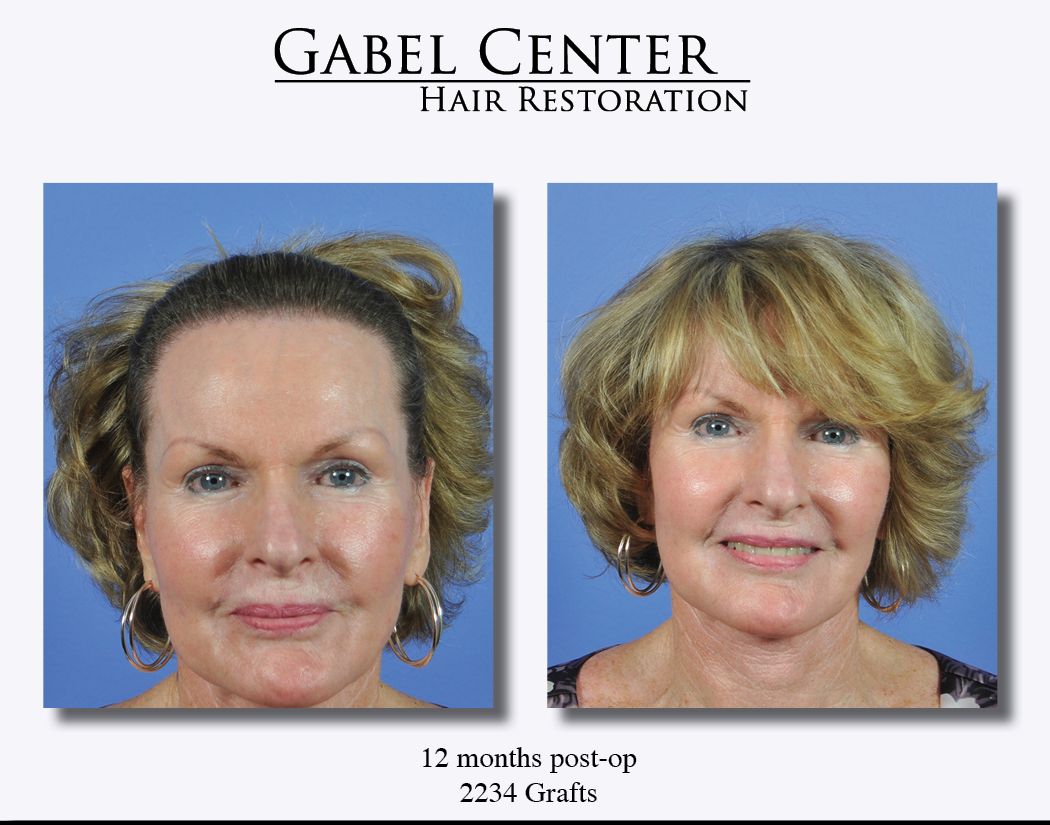
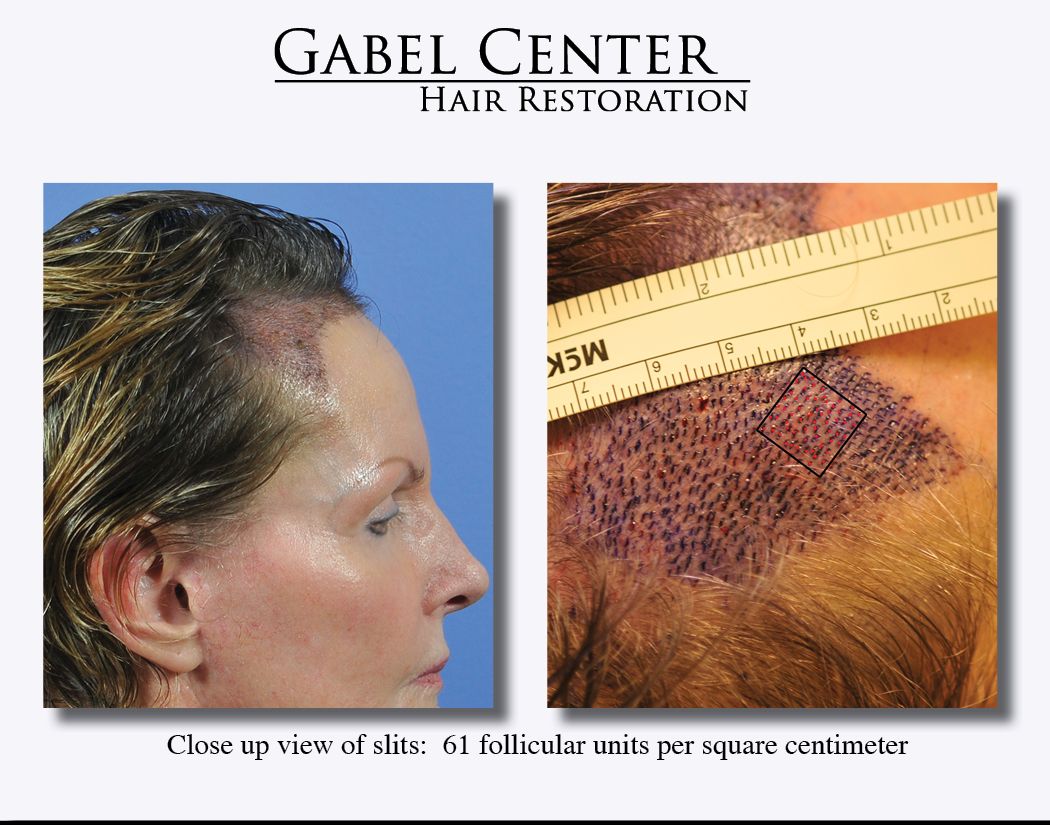
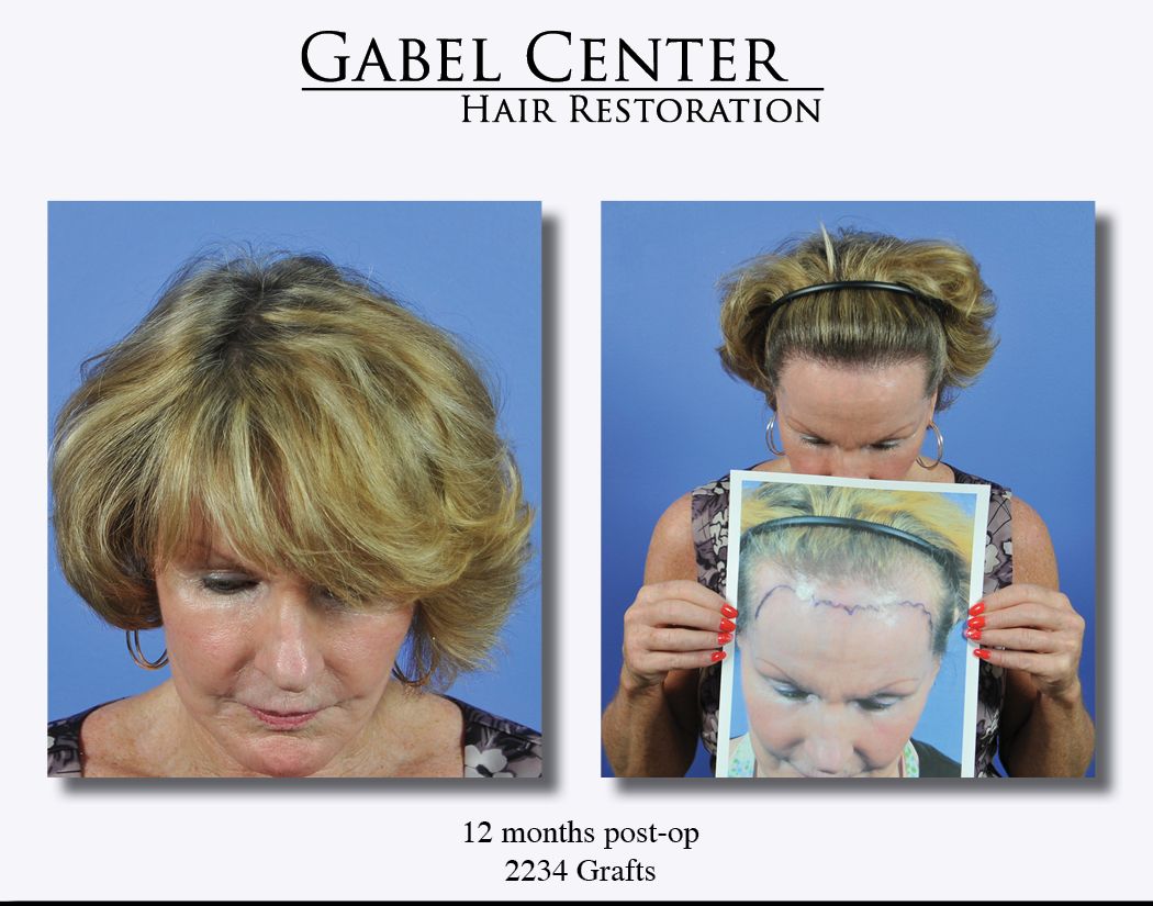
For several years, this patient has noticed a gradual thinning and decline of her frontal hairline. She felt that it was thinning to the point where she was unable to style it to her liking and always had to brush it forward to hide her hairline. It became very frustrating to her and ultimately she was self conscious about it.
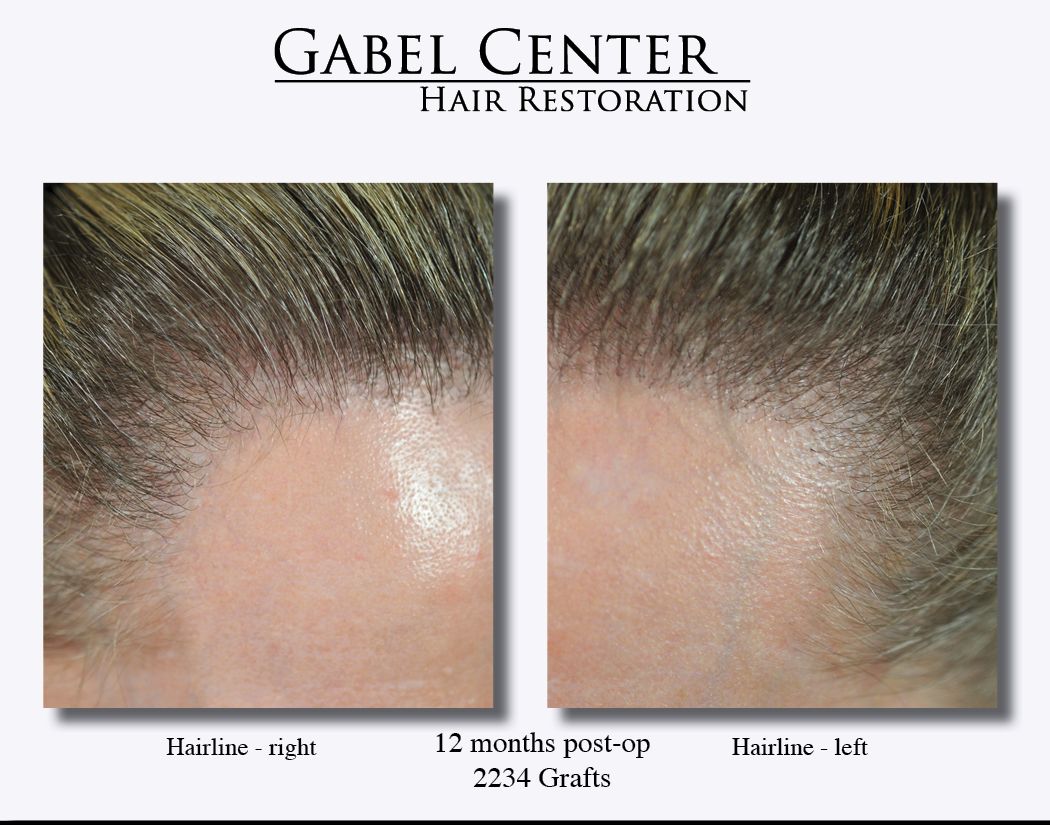
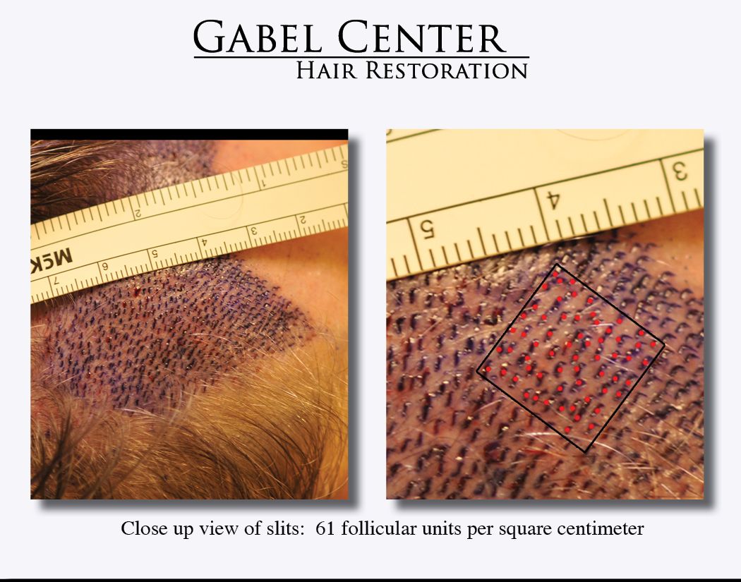
We designed a female pattern hairline by following her current hairline and lowering it to make a very natural female hairline. 2234 grafts were placed into the frontal hairline and immediately behind it to connect to her native hairline. Over 750 single haired grafts were placed just in the immediate 1 - 2 centimeters and the rest of the grafts were used to connect the new hairline to her native hairline. Close up photographs show the density of the frontal hairline around 60 FU/sq. cm. Once the grafts were placed, I surveyed the entire transplanted region, especially the frontal hairline, and additional grafts were placed in any spaces that could accommodate the hair.
Photographs were submitted with similar orientations showing the timeline for the restoration process: immediately after surgery, 1 week, 3 months, and 1 year following the hair transplant procedure. A video demonstrates the 1 year result at approximately 85% growth; we expect the transplanted hairs to mature and thicken up over the next year of growth.
-
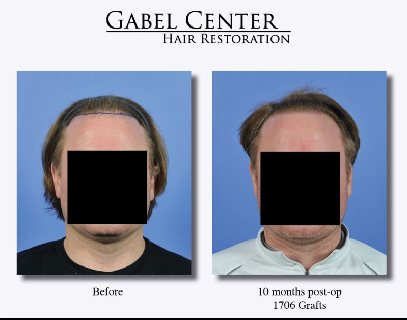
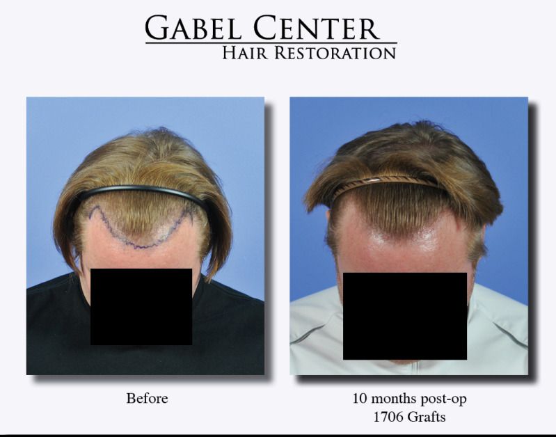
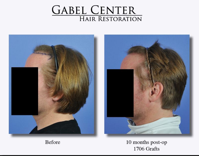
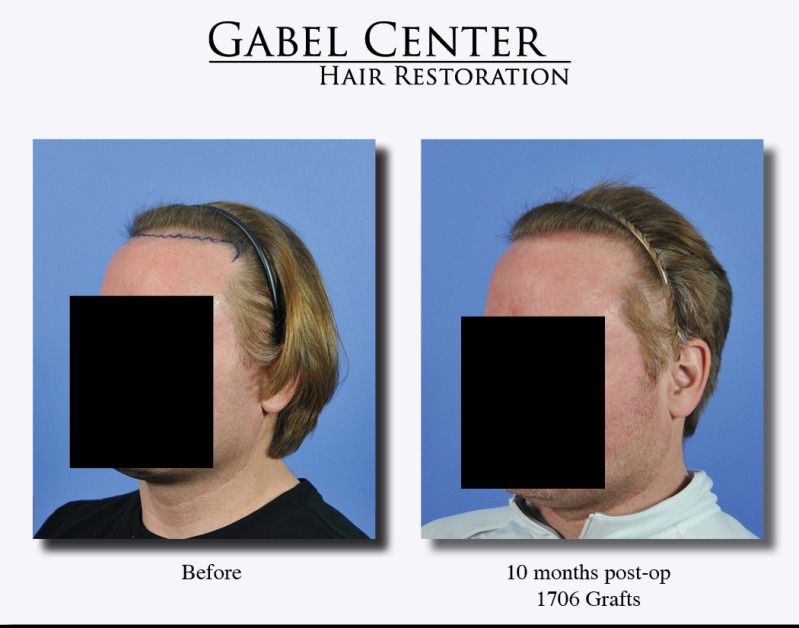
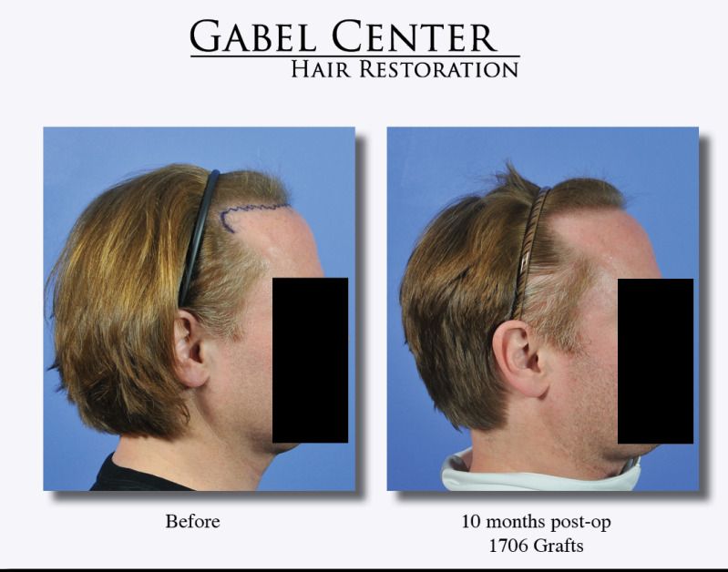
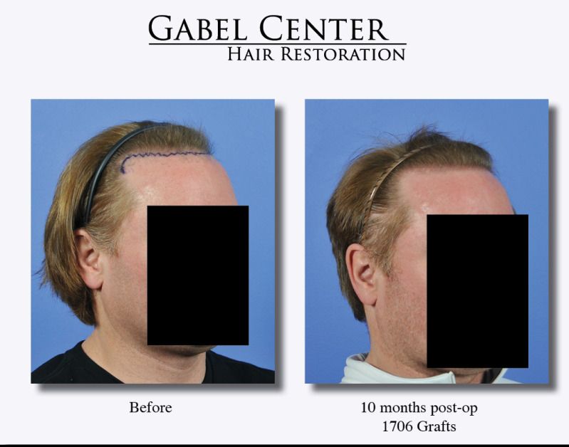
This patient is a 41 year old male who desired to thicken only the immediate frontal hairline. He felt that over the last 12 years that it has very slowly receded and thinned. Although he was concerned about the mid-scalp and crown, this was not an issue at this time. He is now using medical therapy to slow down future hair loss.
1706 grafts were placed in the frontal hairline utilizing high density placement techniques in the coronal plane. The postoperative photographs are 10 months following he procedure.
-
Here are the requested photos. This patient is from out of town so I only have the 1 year result photos. At the time of these photos, he had grown in very well and hopefully I can obtain more recent photographs.


 IMG]
IMG]


Dr. Steven Gabel patient – 15 months out with 2210 grafts, frontal region
in Results Posted by Leading Hair Restoration Clinics
Posted
This patient is a 37-year-old gentleman who is now 15 months out after having 2210 grafts transplanted to the frontal region of the scalp. Since the last visit, he feels the hairs are thickening and maturing.
Overall he is extremely happy with the results. He is able to trim his hair pretty short as seen in the photos and it still has a very natural, full appearance. He is also able to cut the donor area hair down without any evidence of the donor scar showing.